Biochemistry

Biochemistry explores the chemical and molecular basis of life. It includes topics like metabolism, enzymes, proteins, carbohydrates, lipids, and nucleic acids. This subject forms the core of understanding disease mechanisms and drug actions in healthcare.

Biochemistry

CALCIUM

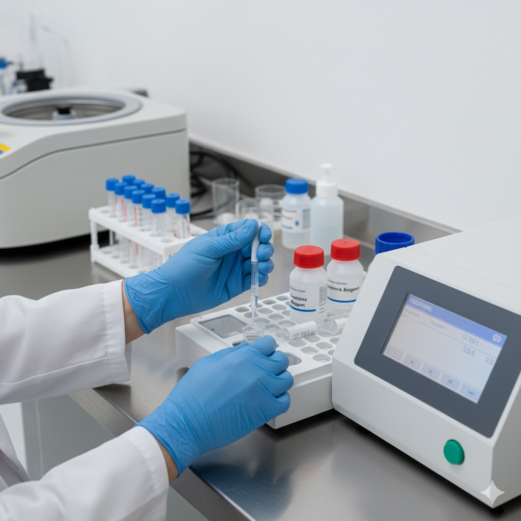
CALCIUM (Arsenazo III) TEST KIT (MONOTEST)INTENDED USE:Intended for in vitro quantitative determination of Calcium in Serum. PRINCIPLE Calcium forms a purple complex with Arsenazo III at neutral pH. Intensity of colour is proportional to calcium concentration and is measured photometrically. Calcium + Arsenazo III → Blue-purple complex CONTENTS Reagent 1: Calcium (ASIII) Reagent – 2 x 15 mL Reagent 2: Calcium Standard – 1 x 1 mL      SAMPLE COLLECTION & PRESERVATION Use serum. Calcium is stable 7 days at 2–8°C when stored in tightly closed glass vials. Avoid haemolysed specimens. REAGENT PREPARATION & STORAGE Store at 2–8°C. Stability up to expiry date when stored properly. Protect from direct light. Do not freeze. PROCEDURE Label test tubes: Blank (B), Standard (S), Sample (T) Add reagents: Reagent 1: 1000 µL to all Standard: 10 µL to S Sample: 10 µL to T Mix and incubate for 5 minutes at 37°C Measure absorbance of S and T against reagent blank at 650 nm  CALCULATION Calcium (mg/dL) = (Absorbance of Test / Absorbance of Standard) × 10 NORMAL VALUES  Serum calcium: 8.5 – 10.5 mg/dL (Values may vary by population and methodology) CLINICAL SIGNIFICANCE Calcium is found mainly in the bones (approx. 98%). In serum, calcium exists in three forms: Protein-bound Ionized (physiologically active) Complexed with anions Serum calcium levels may increase in: Hyperparathyroidism Hyperthyroidism Excess Vitamin D Malignancy Levels may decrease in: Hypoparathyroidism Renal failure Vitamin D deficiency Pancreatitis GENERAL SYSTEM PARAMETERS Reaction type: End point Wavelength: 650 nm (600–660 nm) Reaction temperature: 37°C Sample volume: 10 µL Reagent volume: 1000 µL Standard: 1 mmol/L Incubation: 5 minutes Read against reagent blank LINEARITY Linear up to 15 mg/dL.If sample >15 mg/dL, dilute and multiply result by dilution factor. NOTE Reagent is very stable; colour intensity develops uniformly. Avoid contaminated glassware. LABELING Code No. 317, Pack Size 2 × 15 mL, Standard 1 mL

Biochemistry, Uncategorized

CERATININE

CREATININE REAGENT KIT (Alkaline Picrate Method) INTENDED USE   This reagent kit is used for the in vitro quantitative determination of creatinine in serum and urine. SUMMARY Creatinine is the end product of creatine phosphate metabolism in muscles. It is excreted in urine.Serum creatinine level helps assess kidney (renal) function.Elevated creatinine indicates renal impairment, muscle damage, or muscular dystrophy. PRINCIPLE Creatinine reacts with alkaline picrate to form an orange-colored complex.The intensity of the color is directly proportional to the amount of creatinine present and is measured spectrophotometrically at 520 nm. Reaction:Creatinine + Alkaline Picrate → Orange-Coloured Complex CONTENTS Reagent 1: Creatinine Buffer Reagent Reagent 2: Creatinine Standard Reagent Reagent 3: Creatinine Picrate Reagent MATERIALS REQUIRED  Clean dry glassware Micropipettes & tips Colorimeter or Spectrophotometer Test tubes SPECIMEN Use serum or urine. Serum should be diluted (1:100) in saline. PREPARATION OF REAGENT & STABILITY Reagents are supplied ready-to-use. Store at 2–8°C. Working reagent is stable for 7 days at room temperature (R.T.) or 1 month at 2–8°C. PROCEDURE Pipette into Test Tubes Blank Standard Test Working Reagent 1.0 ml 1.0 ml 1.0 ml Standard – 0.1 ml – Sample – – 0.1 ml Mix well and read absorbance (A) of the Standard and Test after 30 seconds and again after 90 seconds at 520 nm. Distilled water is used as Blank. CALCULATION Creatinine (mg/dL)=ATAS×2text{Creatinine (mg/dL)} = frac{A_T}{A_S} times 2 Creatinine (mg/dL)=ASAT×2 Urine Creatinine (g/L)=ATAS×2×Dilution factortext{Urine Creatinine (g/L)} = frac{A_T}{A_S} times 2 times text{Dilution factor} Urine Creatinine (g/L)=ASAT×2×Dilution factor Where:Aₛ = Absorbance of StandardAₜ = Absorbance of Test NORMAL VALUES Serum: Male – 0.6–1.4 mg/dL; Female – 0.6–1.2 mg/dL Urine (24 hours): 1.0–2.0 g/24 hr (Note: Values may vary by method and laboratory.)                                                  QUALITY CONTROL Use normal and abnormal serum controls to check assay performance. LIMITATION & PRECAUTIONS Avoid hemolysed samples. Maintain precise timing and temperature. Do not use reagents after expiry date. For in vitro diagnostic use only.

Biochemistry

TOTAL PROTEIN

TOTAL PROTEIN (Biuret Method) INTENDED USE:The reagent kit is intended for in vitro quantitative determination of Total Protein in serum/plasma. CLINICAL SIGNIFICANCE:Proteins are conjugations of amino acids, enzymes, hormones and several other kinds of molecules, structural entities in the body. They are involved in the maintenance of the normal distribution of water between blood and the tissues. Considering many of albumin and globulin in the blood has many diagnostic and analytical values as several fractions are involved in many (diseases). Decreased levels are found mainly in malnutrition, hepatic synthesis, protein losses as in hemorrhage or excessive protein catabolism. PRINCIPLE:Proteins, in an alkaline medium, bind with the cupric ions present in the biuret reagent to form a blue-violet colored complex. The intensity of the color formed is directly proportional to the amount of proteins present in the sample. REACTION:Total Protein + Cu²⁺ → Violet complex. CONTENTS:Reagent 1: Biuret ReagentReagent 2: Protein Standard 6 g/dl MATERIALS REQUIRED BUT NOT PROVIDED: Clean Dry Glassware Laboratory Glass Pipettes or Micropipettes & Tips Colorimeter or Bio-Chemistry Analyzer SAMPLES:Serum, Heparinized/EDTA Plasma. Proteins are reported to be stable in the sample for 6 days at 2–8°C. PREPARATION OF REAGENT & STABILITY:All reagents are stable till the expiry date mentioned on the label at standard room temperature.If stored at 2–8°C, this is stable till expiry when not in use.All reagents are ready to use form. GENERAL SYSTEM PARAMETERS: Wavelength: 546 nm (530–570 nm) Temperature: 25°C Light Path: 1 cm Assay Type: End Point Reaction Time: 5 min Standard Concentration: 6 g/dl Zero setting: Reagent Blank Sample Volume: 10 µl Reagent Volume: 1.0 ml PROCEDURE:Pipette into clean dry test tube labeled as Blank (B), Standard (S) and Test (T): Addition sequence Blank (B) Standard (S) Test (T) Biuret Reagent 1.0 ml 1.0 ml 1.0 ml Distilled Water 10 µl — — Standard — 10 µl — Sample — — 10 µl Mix and incubate for 5 minutes at Room Temperature. Measure the absorbance of Standard and Sample (Aₛ) against the Blank (Aᵦ) at 546 nm. CALCULATION:Total Protein Conc. (g/dl) =(Aₛ / Aₛₜ) × 6 NORMAL VALUE:Serum: 6.0–8.0 g/dlIt is recommended that each laboratory establish its own normal range. LINEARITY:This procedure is linear up to 10 g/dl. Samples above this concentration should be suitably diluted and results should be multiplied by dilution factor. QUALITY CONTROL:For accuracy it is necessary to run known controls with every assay. LIMITATION & PRECAUTIONS: Storage condition mentioned on the kit must be adhered to. Do not mix reagents from different lot numbers. Maintain clean dry glassware for accurate results. Samples showing hemolysis are to be avoided. Avoid contamination of reagents with even traces of saliva (as causes false high absorbance due to dirt). BIBLIOGRAPHY: Henry, T.R., Clin. Chem., Am. J. Clin. Path., 16:40 Henry, T.R., Cannon, D.C., Winkleman, J.W., Clinical Chemistry, Principles and Techniques, Harper & Row, 2nd Edition, 1974. CODE NO / PACK SIZE / REAGENT 1 / REAGENT 2217 / 1 × 100 ml / 1 × 5 ml217A / 1 × 50 ml / 1 × 3.0 ml

Biochemistry, Uncategorized

SERUM ALBUMIN

SUMMARY: Albumin a major plasma protein is synthesized in the liver from amino acids, which are absorbed from the liver. Its function includes regulation of distribution of extracellular fluid, transportation of various hormones, steroids, and amino acids. Aim: Estimation of Serum Albumin by BCG (Bromocresol Green) method. PRINCIPLE: Albumin binds with bromocresol green at pH 4.2 causing a shift in absorbance maximum of the Yellow BCG dye. The resulting bluish-green color is measured photometrically. The intensity of the color is directly proportional to the albumin concentration. The absorbance of the test and standard are measured against blank at 630 nm wavelength. Albumin + BCG → Albumin-BCG complex (blue-green color) REQUIREMENTS: Three test tubes Colorimeter with Automatic Pipetter Total Protein working reagent Distilled water Cuvette Pipette Serum Sample PROCEDURE: Contents Blank (ml) Standard (ml) Test (ml) Working reagent 1.0 1.0 1.0 Distilled water 1.0 – – Standard – 0.1 – Serum – – 0.1 For further help WhatsApp us: +91-9891068072 INSTRUCTIONS: Measure all content according to the chart in the test tubes. Mix well and incubate for 5 minutes at 37°C temperature in incubator. Read the optical density of the test and standard at 630 nm wavelength. Calculation: Serum Albumin (g/dL)= (Optical Density of Test × Concentration of Standard) / (Optical Density of Standard) Example Calculation: After testing:Optical Density of Test = 0.318Optical Density of Standard = 0.24 Therefore:= (0.318 × 4.0) / 0.24= 5.3 g/dL Normal Values: Parameter Normal Range Total Protein 6.0 – 8.0 g/dL Albumin 3.5 – 5.0 g/dL Globulin 2.5 – 3.5 g/dL A/G Ratio 1.0 – 2.0 Clinical Significance: Increased levels of albumin are present in cases of dehydration, especially noted for newborns.Decreased levels of albumin are present in conditions like malnutrition, nephrotic syndrome, hepatic

Biochemistry, Uncategorized

UREA

UREA (NED METHOD):- The reagent set is intended for in vitro Quantitative determination of Urea in Serum and plasma. CLINICAL SIGNIFICANCE:Urea is the end product of the protein metabolism. It is synthesized in the liver from ammonia produced by the deamination of amino acids. It is transported by blood to the kidneys where it is excreted. Increased values are found in renal failure, urinary tract obstruction, shock, congestive heart failure and burns. Decreased levels are found in liver failure and pregnancy. PRINCIPLE:Urea forms with ortho–phthalaldehyde and Naphthylethylenediamine in the acidic medium a coloured complex. The value of colour formed is directly proportional to the urea concentration in the test sample and is measured by a fixed time method at 550 nm. REACTION:Urea + ODA → NH₃ + H₂ONH₃ + NED → Orange Color Complex. CONTENTS:Reagent 1 : ODA ReagentReagent 2 : NED ReagentReagent 3 : Urea Standard, 50 mg/dl MATERIALS REQUIRED BUT NOT PROVIDED: Clean Dry Glassware Laboratory Glass Pipettes or Micropipettes & Tips Bio–Chemistry Analyse SAMPLES: Serum, Heparinized/EDTA Plasma. Urea is reported to be stable in the serum for 5 days when stored at 2–8°C. PREPARATION OF REAGENT & STABILITY:All the reagents are ready for use and stable till the expiry date mentioned on the label when stored at 2–8°C. GENERAL SYSTEM PARAMETERS:Reaction type: End pointWavelength: 550 nm (520–550 nm) (Increasing)Temperature: 37°CReagent Volume: 1.0 mlSample Volume: 10 µlZero setting: Against Reagent BlankLight path: 1 cm. PROCEDURE:Pipette into clean dry test tubes labeled as Standard (S) and Test (T) Addition sequence (S) (T) ODA Reagent 1.0 ml 1.0 ml Sample — 10 µl Standard 10 µl — Mix well and incubate at 37°C for 5 minutes NED Reagent 0.05 ml 0.05 ml Mix well and read the absorbance A₁ of the standard and test after exactly 5 minutes. Read A₂ after exactly 10 minutes. The absorbance reading to be recorded at 550 nm.Finally, take the difference A₂–A₁ for both the standard and test. For Standard: ΔA = A₂S – A₁SFor Test: ΔA = A₂T – A₁T CALCULATION:Urea Concentration (mg/dl) =(ΔAT / ΔAS) × 50 NORMAL VALUE:Serum/plasma: 15–40 mg/dlEach laboratory should establish its own normal range depending on the population. LINEARITY:The method is linear upto 200 mg/dl. The value exceeding 200 mg/dl should be diluted appropriately with distilled water and the values obtained multiplied by dilution factor. QUALITY CONTROL:For accuracy it is necessary to run known controls with every assay. LIMITATION & PRECAUTIONS: Storage condition of the reagent and kit should be strictly followed. Avoid contamination of reagents. All glassware must be dry and free from detergent or debris. BIBLIOGRAPHY: Goodwin, J., Hart, T., Am. J. Chem., 26 (1977) 707 CODE NO. PACK SIZE Reagent 1 Reagent 2 Reagent 3 Z18 100 ml 1 x 100 ml 1 x 50 ml 1 x 3.0 ml BEACON DIAGNOSTICS PVT. LTD.424, NEW GIDC, KABILPORE, NAVSARI – 396 424, INDIA

Scroll to Top