
Clinical Pathology

Clinical Pathology deals with the examination of body fluids like blood, urine, and CSF. It helps diagnose diseases through chemical, hematological, and microscopic tests. A key category for lab technicians and diagnostic professionals.

Clinical Pathology

Cerebrospinal fluid (CSF) examination

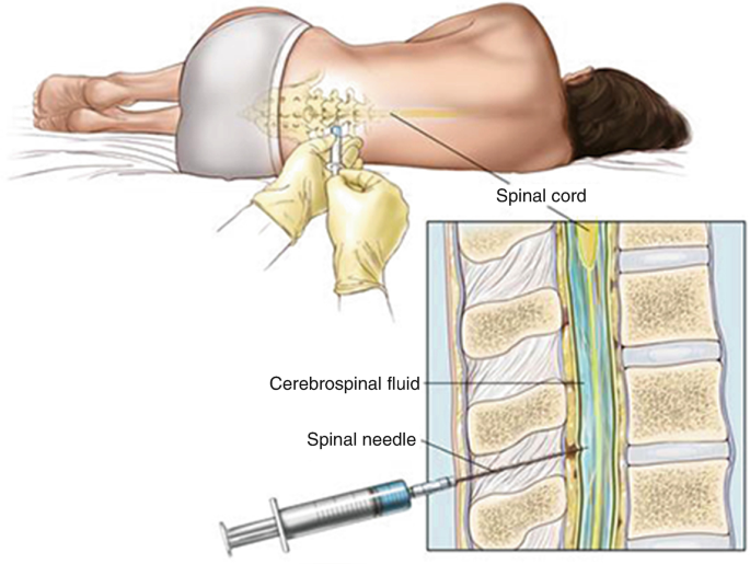
Cerebrospinal fluid(CSF):- CSF are produced by the choroid plexus of the lateral, third, and fourth ventricles up to 80% approximetly and 20% CSF are produced by the surface of brain and spinal card. CSF is a clear, colorless, sterile fluid that fills the ventricles of the brain, the central canal of the spinal cord, and the subarachnoid space surrounding the brain and spinal cord.  CSF produced about 500 mL per day, although only about 150 mL is present at any given time because it is continuously reabsorbed into the bloodstream through the arachnoid villi and granulations. CSF circulates through the ventricular system and around the central nervous system, forming a protective fluid environment for neural tissues. CSF performs several essential physiological functions. It acts as a mechanical cushion,  shocks absorbing and protecting the delicate brain and spinal cord from trauma. It provides buoyancy, reducing the effective weight of the brain.CSF also helps maintain chemical homeostasis The CSF examination or dignosis is important for diagnose infections such as Meningitis and Encephalitis, inflammatory disorders such as Multiple Sclerosis, hemorrhages, malignancies, and other neurological conditions. CSF acts as a mechanical cushion for the brain and spinal cord, absorbing shocks and protecting them from injury caused by sudden movements or impacts. CSF collection :- SF collection is the procedure of obtaining a sample of cerebrospinal fluid (CSF) for laboratory examination to diagnose diseases.The most common method of CSF collection is lumbar puncture (spinal tap).The lumber puncture is done by expert physician , surgeon & Nurses. Procedure for CSF collection:- 1.The patient is usually placed in a lateral decubitus (side-lying) or sitting position. 2.Cleaning the skin and administering local anesthesia. 3.A sterile spinal needle is inserted into the subarachnoid space between the L3–L4 or L4–L5 vertebrae,below the end of     the spinal cord. 4.The needle enters the subarachnoid space, CSF flows through the needle and is collected into sterile tubes. 5.Specimen centrifuged. then the supernatant part used for biochemical tests and the sediment is used for weight mount preparation ,gram’s staining , etc. Physical examination of CSF:– It consist of following examination:- 1.Odour:- CSF are odourless normally in few of bacterial infection conditions putrified small occures. 2. Colour:- Colourless but in bacterial menengitis & physical damage causes redish colour CSF. 3.pH:- Normally pH of CSF is 7.35 – 7.4. 4.Volume:- In adults 120-150 ml CSF are flow in around the spinal card & brain,In child 80-120 ml CSF and neonate 10-60 ml.The produced rate of CSF is 500 ml per day. 5.Specific gravity:- Normal specific gravity  1.003-1.008. 6.Appearence:- Normally CSF are clear but in bacterial menengitis. its may cloudly or Frankly purulent.(Frankly- clear or clean, purulent-containing pus.). Chemical examination:- CSF Sugar GOD/POD Methods:-  Aim:– Performed CSF sugar test by GOD/POD (Glucose oxidase & peroxydase) method. Principle:-      1.      Glucose + H₂O      Glucose Oxidase           Gluconic acid +H₂O₂     2.      H₂O₂  +   4AAP(Aminoantipyrine) + Phenol    Peroxidase       Quinoneimine dye. 1. Take a three clean and dry test tube and mark as blank standerd and test. 2.Measures all contents according to chart. 3.Mixwell and incubate for 10 minutes at 37 degree celcious temperatures. 4. Read the optical density of the test and standered against blank.at500-520 nm wave length.  Calculation:- CSF glucose calculation =               OD of test               ×  Concentration of Standard                                                                                                                                                                                                 OD of standered Concentration of Standard       = 100 mg/dl Result:-…………….? Normal Value:-                                60-80 mg/dl Clinical significance:- Decreased CSF Glucose (Hypoglycorrhachia):-     Microorganisms and inflammatory cells consume glucose, lowering its concentration in CSF in condition      Bacterial meningitis,tuberculous meningitis, fungal meningitis , malignant infiltration of the meninges,severe CNS inflammation. Increased CSF glucose(hyperglycemia):- CSF glucose generally rises in proportion to blood glucose levels.hyperglycemia (Diabetes mellitus).                                            -:CSF Microprotein:- Aim:-  Performed CSF protein by biurate methods. Principle:-  CSF protein + Cu2    Alkaline medium      Violet color complex (Cu-Protein Complex) Requirment:- Test tube, micro-pepette,D/W , calorimeter, CSF sample, working reagen , incubator, tissue paper. 1. Take three clean and dry test tube and mark as blank standerd and test. 2.Measures all contents according to chart. 3.Mixwell and incubate for 5 minutes at 37 degree celcious temperatures. 4. After incbation measure and read the optical density of the test and standered against blank at 540 nm wave length. Calculation:- CSF protein =               OD of test      ×  Concentration of Standard                                                                                                                                                                                                         OD of standered Concentration of Standard    = 6 mg/dl Result:-.………………………? Normal value:-               15-45 mg/dl Clinical significance:- CSF microprotein estimation helps diagnose neurological disorders. Increased CSF protein is observed in meningitis, tuberculous
Clinical Pathology

SEMEN ANALYSIS PRACTICALE

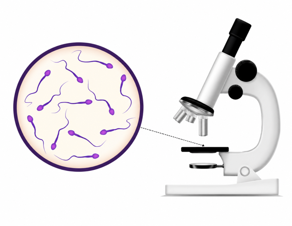
Semen analysis:- Semen analysis is performed to evoluvate male firtility and reproductive health assessing the quality and quantity of sperm in a semen sample. Semen examination is an inexpensive test used for the infertility ,success of vasectomy and medicolegal  cases,hormonal imbalances, infections, lifestyle factors, or exposure to certain medications and environmental toxins. semen analysis consist of following steps. 1.Rutine analysis:- Routine Analysis of Semen is a laboratory test performed to evaluate the physical, microscopic, and functional characteristics of semen. The examination includes assessment of semen volume, color, viscosity, liquefaction time, pH, sperm concentration (count), motility, morphology , and the presence of other cells such as white blood cells or immature germ cells. process included:- 1.Sample collection 2.physical examination  3.Chemical examination 4.Microscopic examination  1.Sample collections:-  A semen specimen should be collected after 3-7 days  of sexual absentees for accurate results.The sample is usually obtained by masturbation directly, the specimen is collected in a clean, sterile, wide-mouth ,leak-proof container provided by the laboratory or properly washed dry condom.then specimen submitted in a laboratory. After collection, the specimen container properly labeled with the patient,s name ,date ,and time of collection. sample should be allowed to liquefy at room tempreture. 2.Physical examination or Gross examination:- The physical examination of semen is the first step in routine semen analysis.its consist following aspect. 1.Volume:- Volume in a single ejaculation approximately 2.5-5 ml semen passed ,the volume is slightly more in patient of infertility. The volume dose not various with the periods of absenties in increase volume of semen is known as hyperspermia and low  volume is caused hypospermia. 2.colour:-Normally it is whitish ,gray white of slightly yellowish or milky.Yellowish or reddish discoloration may indicate infection or blood contamination. 3.pH(power of hydrogen ):- The normal pH  is 7-8.it is slightly alkaline in nature. 4.Viscosity :- During the ejaculation semen are viscus ,when ejaculation falls drop by drop . 5.Liquification:- Liquification occur because of presence of fibrinogen ,normally liquification occur at room tempreature within 10-30 minute avarage 20 minutes. 2. Chemical examination:- Fructose test of semen:- Fructose test is used to determined androgen deficiency or ejaculatory obstruction to semen .the level of seminal fructose is low in both condition fructose is measured qualitative by resorcinol’s. The fructose test is performed to detect the presence and amount of fructose in seminal fluid. Fructose is produced mainly by seminal vesicles and serves as the primary energy source for sperm motility. Requirements:-  *Diluted HCL *Resorcinol *Semen/specimen *Test tube  *Bansal burner  Procedure:- 1.  Take a 5 ml diluted HCL in a test tube. 2.  Add 1ml semen. 3. Add 5 mg resorcinol’s boil it for few minuts. Normal value:-                                        150-600 mg/dl. Interpretation:- Appearence of red colour indicate the presence of fructose. it can measured by spectrophotometer.                             -:Microscopic examination:- Semen examine under microscope motility of spermatozoa counting & spermtozoa morphology. Motility:- Motility with the help of liquefied semen make a wet mount sample slide and examin under the microscope first low power field and then highpowerr field microscope .normally within 2 hours of ejaculation mote than >60% spermatozoa  vagrasly motile, and in 6-8 hours 25-40% are motile.                                -:Spermatozoa counting:- Aim:- Spermatozoa counting done by improves new baur’s chamber. Principle:- Diluted spermatozoa charged in new baur’s chamber and count the 4 square cornor. Requirment :-  *Improved new baur’s chamber *Micro pepette*Semen*Diluting fluid*Test tube*Coverslips Diluting fluid:-  Sodium bicarbonate                    5 gmFormaline                                     10%Distilled water                             100 ml. Peocedure:- 1.Dilute the semen specimen in 1:10. 2.Charge the new baur’s chamber and weight for 2 minutes to allow the spermatozoa settle down. 3.Examine under microscope and count spermatozoa in 4 square cornor of new baur’s chamber and calculate them as follow. Calculation :-   Spermatozoa =  N×D/V =N×10/0.4 =N×25 Spermatozoa /ml= N×25×1000 =N×25000 Normal values:-     >60 million/ml >60 × 10⁶/ml Abnormal values:- <20 × 10⁶/ml Marphology examination of spermatozoa:- For the morphology’s  study of spermatozoa prpared a thin smear from liquified semen on a glass slide,and stained it with any of the romanowashky stain,pap stain or H&E stain. Observe atleast 200 spermatozoa for any abnormalities in their morphology normally 80% spermatozoa are normal. Normal marphology of spermatozoa:-Abnormal marphology of specimen. spermatozoa are motile reproductive cells a smooth rimmed oval shaped  head with 2.5  –  3.5µm wide and 5-6 µm  longan uncoilled 45 uncoild 45 µm long tail that should be thiner than head and mid piece.midpiece of the spermatozoa is a sigment between the head & mid piece is approximatlh similor to the head  Abnormal marphology of specimen:- Abnormal spermatozoa are aneble to fertilized egg. Abnormaltise in spermatozoz head:- 1.Detached or hatched head.The head of the sperm has separated from its tail. Such sperm are generally non-functional and cannot swim properly. 2.Large head:-A large-headed spermatozoon (also called macrocephalic sperm) is a sperm cell whose head is larger than normal. 3.Small head :-A sperm with a smaller-than-normal head, often associated with abnormal DNA/chromosome content and reduced fertility potential. 4.Elongated head:-A sperm with an abnormally long, narrow head. 5.Irregular head.A sperm with an abnormally shaped head; it is a morphology defect that may impair fertilization ability. 6.Amorphous head ;-A sperm with a head that lacks a normal. 7.Small acrosome head:-A sperm with an abnormally small acrosome (the cap-like structure on the head) 8.No acrosome head.:- A sperm lacking the acrosome on its head. Abnormal neck or mid poece defect:– Asemetric neck:-sperm in which the neck (connecting piece between the head and midpiece) is attached off-center or at an abnormal angle to the head. Bent neck :- A sperm in which the neck is sharply bent relative to the head or midpiece. Thin neck:-A

Clinical Pathology

MEN INFERTILITY

MEN INFERTILITY:- Male infertility is any health issue in a man that lowers the chances of his female partner getting pregnant. About 13 out of 100 couples can’t get pregnant with unprotected sex. There are many causes for infertility in men and women. In over a third of infertility cases, the problem is with the man. This is most often due to problems with his sperm production or with sperm delivery. What Happens Under Normal Conditions? The man’s body makes tiny cells called sperm. During sex,ejaculation normally delivers the sperm into the woman’s body. The male reproductive system makes, stores, and transports sperm. Chemicals in your body called hormones control this. Sperm and male sex hormone (testosterone) are made in the 2 testicles. The testicles are in the scrotum, a sac of skin below the penis. When the sperm leave the testicles, they go into a tube behind each testicle. This tube is called the epididymis. Just before ejaculation, the sperm go from the epididymis into another set of tubes. These tubes are called the vas deferens. Each vasdeferens leads from the epididymis to behind your bladder in the pelvis. There each vas deferens joins the ejaculatory duct from the seminal vesicle. When you ejaculate, the sperm mix with fluid from the prostate and seminal vesicles.This forms semen. Semen then travels through the urethra and out of the penis. Male fertility depends on your body making normal sperm and delivering them. The sperm go into the female partner’s vagina. The sperm travel through her cervix into her uterus to her fallopian tubes. There, if sperm and egg meet, fertilization happens. Causes:– Making mature, healthy sperm that can travel depends on many things. Problems can stop cells from growing into sperm. Problems can keep the sperm from reaching the egg. Even the temperature of the scrotum may affect fertility. These are the main causes of male infertility: Sperm Disorders Varicoceles Retrograde Ejaculation Immunonologic Infertility Obstruction Hormones Medication Sperm Disorders:- The most common problems are with making and growing sperm. Sperm may: not grow fully be oddly shaped not move the right way be made in very low numbers (oligospermia) not be made at all (azoospermia) Sperm problems can be from traits you’re born with. Lifestyle choices can lower sperm numbers. Smoking, drinking alcohol, and taking certain medications can lower sperm numbers. Other causes of low sperm numbers include long-term sickness (such as kidney failure), childhood infections (such as mumps), and chromosome or hormone problems (such as low testosterone). Damage to the reproductive system can cause low or no sperm. About 4 out of every 10 men with total lack of sperm (azoospermia) have an obstruction (blockage) within the tubes the sperm travel through. A birth defect or a problem such as an infection can cause a blockage. Varicoceles:-  Varicoceles are swollen veins in the scrotum. They’re found in 16 out of 100 of all men. They are more common in infertile men (40 out of 100). They harm sperm growth by blocking proper blood drainage. It may be that varicoceles cause blood to flow back into your scrotum from your belly. The testicles are then too warm for making sperm. This can cause low sperm numbers. For more information please refer to the Varicoceles information page. Retrograde Ejaculation:- Retrograde ejaculation is when semen goes backwards in the body. They go into your bladder instead of out the penis. This happens when nerves and muscles in your bladder don’t close during orgasm (climax). Semen may have normal sperm, but the semen is not released from the penis, so it cannot reach the vagina. Retrograde ejaculation can be caused by surgery, medications or health problems of the nervous system. Signs are cloudy urine after ejaculation and less fluid or “dry” ejaculation. Immunologic Infertility:– Sometimes a man’s body makes antibodies that attack his own sperm. Antibodies are most often made because of injury, surgery or infection. They keep sperm from moving and working normally. We don’t know yet exactly how antibodies lower fertility. We do know they can make it hard for sperm to swim to the fallopian tube and enter an egg. This is not a common cause of male infertility. Obstruction:- Sometimes the tubes through which sperm travel can be blocked. Repeated infections, surgery (such as vasectomy), swelling or developmental defects can cause blockage. Any part of the male reproductive tract can be blocked. With a blockage, sperm from the testicles can’t leave the body during ejaculation. Hormones:- Hormones made by the pituitary gland tell the testicles to make sperm. Very low hormone levels cause poor sperm growth. Chromosomes:- Sperm carry half of the DNA to the egg. Changes in the number and structure of chromosomes can affect fertility. For example, the male Y chromosome may be missing parts. Medication:- Certain medications can change sperm production, function and delivery. These medications are most often given to treat health problems like: arthritis depression digestive problems anxiety or depression infections high blood pressure cancer Diagnosis:- Causes of male fertility can be hard to diagnose. The problems are most often with sperm production or delivery. Diagnosis starts with a full history and physical exam. Your health care provider may also want to do blood work and semen tests. History and Physical Exam:- Your health care provider will take your health and surgical histories. Your provider will want to know about anything that might lower your fertility. These might include defects in your reproductive system, low hormone levels, sickness or accidents. Your provider will ask about childhood illnesses, current health problems, or medications that might harm sperm production. Such things as mumps, diabetes and steroids may affect fertility. Your provider will also ask about your use of alcohol, tobacco, marijuana and other recreational drugs. He or she will ask if you’ve been exposed to radiation, heavy metals or pesticides. Heavy metals are an exposure issue (e.g. mercury, lead arsenic). All of these can affect fertility. Your health care provider will learn how your body works during sex. He or she will want to

Scroll to Top