


Cerebrospinal fluid(CSF):-
CSF are produced by the choroid plexus of the lateral, third, and fourth ventricles up to 80% approximetly and 20% CSF are produced by the surface of brain and spinal card. CSF is a clear, colorless, sterile fluid that fills the ventricles of the brain, the central canal of the spinal cord, and the subarachnoid space surrounding the brain and spinal cord.
CSF produced about 500 mL per day, although only about 150 mL is present at any given time because it is continuously reabsorbed into the bloodstream through the arachnoid villi and granulations.
CSF circulates through the ventricular system and around the central nervous system, forming a protective fluid environment for neural tissues.
CSF performs several essential physiological functions. It acts as a mechanical cushion, shocks absorbing and protecting the delicate brain and spinal cord from trauma.
It provides buoyancy, reducing the effective weight of the brain.CSF also helps maintain chemical homeostasis
The CSF examination or dignosis is important for diagnose infections such as Meningitis and Encephalitis, inflammatory disorders such as Multiple Sclerosis, hemorrhages, malignancies, and other neurological conditions.
CSF acts as a mechanical cushion for the brain and spinal cord, absorbing shocks and protecting them from injury caused by sudden movements or impacts.
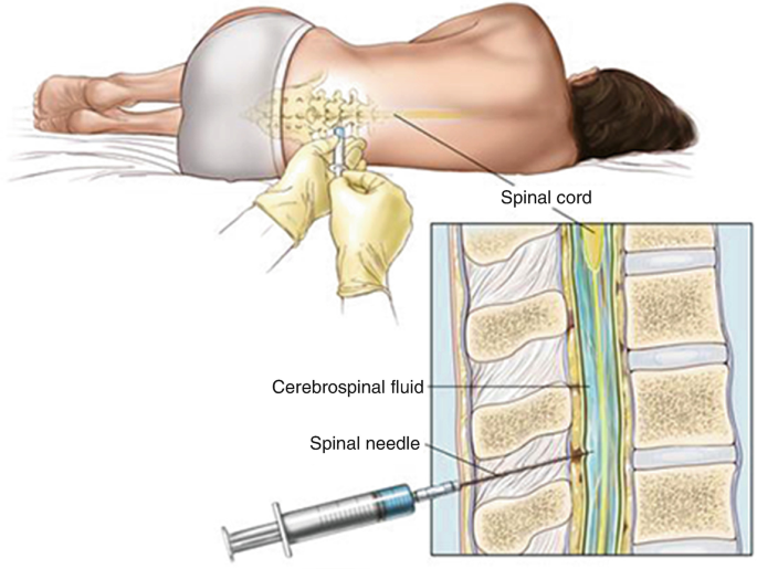
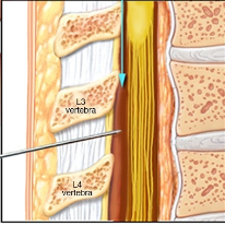
CSF collection :-
The lumber puncture is done by expert physician , surgeon & Nurses.
5.Specimen centrifuged. then the supernatant part used for biochemical tests and the sediment is used for weight mount preparation ,gram’s staining , etc.
Physical examination of CSF:– It consist of following examination:-
1.Odour:- CSF are odourless normally in few of bacterial infection conditions putrified small occures.
2. Colour:- Colourless but in bacterial menengitis & physical damage causes redish colour CSF.
3.pH:- Normally pH of CSF is 7.35 – 7.4.
4.Volume:- In adults 120-150 ml CSF are flow in around the spinal card & brain,In child 80-120 ml CSF and neonate 10-60 ml.The produced rate of CSF is 500 ml per day.
5.Specific gravity:- Normal specific gravity 1.003-1.008.
6.Appearence:- Normally CSF are clear but in bacterial menengitis. its may cloudly or Frankly purulent.(Frankly- clear or clean, purulent-containing pus.).
Chemical examination:-
CSF Sugar GOD/POD Methods:-
Aim:– Performed CSF sugar test by GOD/POD (Glucose oxidase & peroxydase) method.
Principle:-
2. H₂O₂ + 4AAP(Aminoantipyrine) + Phenol Peroxidase Quinoneimine dye.
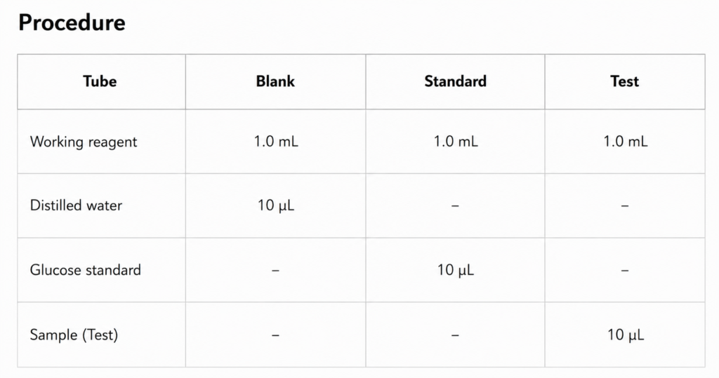
1. Take a three clean and dry test tube and mark as blank standerd and test.
2.Measures all contents according to chart.
3.Mixwell and incubate for 10 minutes at 37 degree celcious temperatures.
4. Read the optical density of the test and standered against blank.at500-520 nm wave length.
Calculation:-
CSF glucose calculation = OD of test × Concentration of Standard OD of standered
Concentration of Standard = 100 mg/dl
Result:-…………….?
Normal Value:- 60-80 mg/dl
Clinical significance:-
Decreased CSF Glucose (Hypoglycorrhachia):-
Bacterial meningitis,tuberculous meningitis, fungal meningitis , malignant infiltration of the meninges,severe CNS inflammation.
Increased CSF glucose(hyperglycemia):-
hyperglycemia (Diabetes mellitus).
-:CSF Microprotein:-
Principle:-
CSF protein + Cu2 Alkaline medium Violet color complex (Cu-Protein Complex)
Requirment:-
Test tube, micro-pepette,D/W , calorimeter, CSF sample, working reagen , incubator, tissue paper.
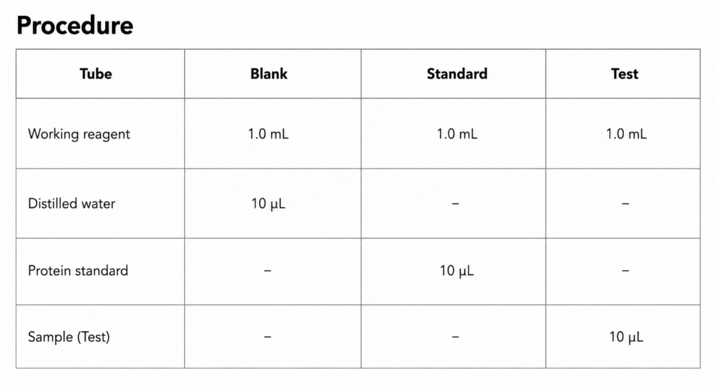
1. Take three clean and dry test tube and mark as blank standerd and test.
2.Measures all contents according to chart.
3.Mixwell and incubate for 5 minutes at 37 degree celcious temperatures.
4. After incbation measure and read the optical density of the test and standered against blank at 540 nm wave length.
Calculation:-
CSF protein = OD of test × Concentration of Standard OD of standered
Concentration of Standard = 6 mg/dl
Result:-.………………………?
Normal value:- 15-45 mg/dl
Clinical significance:-
CSF microprotein estimation helps diagnose neurological disorders. Increased CSF protein is observed in meningitis, tuberculous meningitis, multiple sclerosis, brain tumors, hemorrhage, and spinal cord diseases, whereas decreased protein levels may occur in CSF leakage. Therefore, CSF microprotein measurement is an important tool in the evaluation of central nervous system disorders.
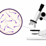
-:Microscopic examination of CSF:-
Aim:- Performed CSF RBCs count by New baur’s chamber.
Principle:- CSF sample diluted with CSF diluting fluid in 1:1 the fluid presence.
Requirment:-
CSF diluting fluid , New baur’s chamber , CSF sample , Micro pepette . Tissue paper , Microscope . Coverslip etc.
Procedure:-
1. For RBCs counting take clean and dry test tube and dilute CSF specimen in 1:1 by adding 9µl diluting fluid and 1µl CSF sample or used for undiluted sample .
2. Make homogeneous suspension and charge the New baur’s chamber.
3. Weight for 2 minutes to settle down the RBCs in chamber.
4. Set the chamber of under high power field of microscope and count cell present in center square 5 out of 25
Calculation:-
RBCs per µL= Number of RBCs counted ×Dilution factor volume counted
Labs usually directly report: RBCs / µL (mm³).
Normal values:- 0 RBCs/µL (mm³)
clincal significance:-“Presence of RBCs in CSF indicates subarachnoid hemorrhage, intracranial hemorrhage, or a traumatic lumbar puncture (traumatic tap). Persistent RBCs in all tubes suggest hemorrhage, while decreasing RBCs in successive tubes suggest a traumatic tap.”
CSF WBCs count:-
Aim:- Performed CSF WBCs count by the New baur’s chamber(hemocytometer).
Principle:- WBCs counting is done by diluting CSF in Newbaour’s chamber with WBC diluting fluid(Trunk’s fluid)which cause lysis of RBCs and stain.
Requirment:-
Newbaur’s chamber ,Micropepette,Coverslip ,Microscope,Diluting fluid, Tissue paper ,Test tube rack, CSF sample etc.
Procedure:-
1.Mix the sample carefully.
2. If the sample is clear , use undiluted ,if sample is cloudy, make 1:20 dilution by using WBC pipette (draw CSF UP TO 0.5 mark and dilute with the CSF diluting fluid (Trunk’s fluid) up to 11 mark.) mix well.
3.Charge the newbaur;s chamber with homogenous diluted CSF and weigth for 2 minutes allow the cell settle down the WBC under low power field in 4 square.
Calculation:-
WBCs/µL = Number of cells counted×Dilution factor Area counted × Depth
or
= Number of cell counted × Dilution factor volume
= N × D V
Area = 9 mm²
Depth = 0.1 mm
Volume = 0.9 mm³ (= 0.9 µL)
Normal value:- Adults: 0–5 WBCs/µL (mm³)
CSF WBCs count:-
Aim:- Performed CSF WBCs count by the New baur’s chamber(hemocytometer).
Principle:- WBCs counting is done by diluting CSF in Newbaour’s chamber with WBC diluting fluid(Trunk’s fluid)which cause lysis of RBCs and stain.
Requirment:-
Newbaur’s chamber ,Micropepette,Coverslip ,Microscope,Diluting fluid, Tissue paper ,Test tube rack, CSF sample etc.
Procedure:-
1.Mix the sample carefully.
2. If the sample is clear , use undiluted ,if sample is cloudy, make 1:20 dilution by using WBC pipette (draw CSF UP TO 0.5 mark and dilute with the CSF diluting fluid (Trunk’s fluid) up to 11 mark.) mix well.
3.Charge the newbaur;s chamber with homogenous diluted CSF and weigth for 2 minutes allow the cell settle down the WBC under low power field in 4 square.
Calculation:-
WBCs/µL = Number of cells counted×Dilution factor Area counted × Depth
or
= Number of cell counted × Dilution factor volume
= N × D V
Area = 9 mm²
Depth = 0.1 mm
Volume = 0.9 mm³ (= 0.9 µL)
Normal value:-
Adults: 0–5 WBCs/µL (mm³)
Newborns: Up to 20–30 WBCs/µL
Clinical significance:-
An increased CSF WBC count (pleocytosis) indicates infection, inflammation, or disease of the central nervous system. Neutrophils are commonly increased in bacterial meningitis, while lymphocytes predominate in viral meningitis and tuberculous meningitis.
Different leukocytes count (DLC):-
Aim:- Examination of peripheral CSF film for different leukocytes count.
Procedure:-
Prepared CSF smear (thin) and stained it which leisman or gimsa or field stain and examination under the microscope as follows:-
1. Choose on area of the smear for microscopic .
2. By smear moving the slide horizental direction under the high power field or oil emersion objective lens and identify the WBCs as pertable.
3. Morphology of cell the start the counting type of WBCs and go entering N or P , Neutrophls or polymorph, lymphocyte , Monocytes, Eosinophils and Basophils count 100 different different WBCs cells.
Types of cells.