Microbiology

Microbiology explores the world of microorganisms such as bacteria, viruses, fungi, and parasites. It is vital for understanding infections, disease transmission, sterilization, and laboratory identification of pathogens.

Microbiology

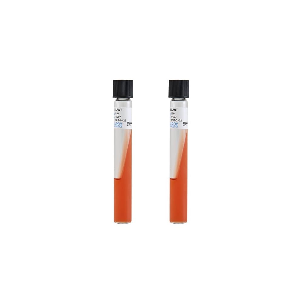
Biochemical Reactions on Triple Sugar Iron Agar (TSI) Slants.

TSI slants are useful in the identification of Enterobacteria by their specific reactions on the slants. v  Principle:- 1.     Alkaline reaction (red color) is shown by the organisms, who fail toferment any one of the sugars. 2.     Fermentation of the sugars is indicated by yellow color since pHrange of phenol red is 6.8 to 8.4 and color change from yellow to red. Sincethe glucose (dextrose) present on the surface of the medium is used up andsince the surface of the slant is exposed to atmosphere, under aerobicconditions, the acid reaction on the surface reverts to alkaline (red color) in18 to 24 hours. (which is a critical duration for this observation). In thebutt, since anaerobic condition exists, the color of the butt remains yellow. 3.     Gas production (carbon dioxide) is indicated by splitting of theagar 4.     Production of hydrogen sulfide imparts black shade to the slant byreacting with ferrous ions. It is an indication of H,S producing organisms. v  Procedure:- 1.      Streak the TSI slant with a loop and stab with a straight needle. 2.      Incubate at 37°C for 18-24 hours.  3.      The various reactions obsrved on the slants are as follows. v Quality control:- 1.      Use the following microorganisms to confirm the reliability of TSIslants. 2.      Positive control:   Acid slantand butt with gas: E. coli. 3.      Negative control:   Alkalineslant and acid butt and no gas: S. paratyphi A.

Microbiology

O-Nitrophenyl-B-D-Galactopyranoside (ONPG) test

O-Nitrophenyl-B-D-Galactopyranoside (ONPG) test:-  v   Principle:- This test is used to detect an organism which produces B-galactosidase enzyme, thathydrolyses ONGP to produce the yellow colored end product orthonitrophenol(ONP). v Requirements:- 1.      ONPG disk 2.      Pure culture of suspected organism 3.      Normal saline v  Procedure:- 1.      Suspend loopful of the organisms in normal saline. 2.      Place an ONPG disk in the tube 3.      Incubate for 4 hours at 37°C. 4.      Observe the tubes for color change  v Observations:- 1             Yellow color of disk: Organism like E. coli 2              No change of color of disk: Negative test v Quality control:- 1.       Use following microorganisms to confirm the reliability of reagents: 2.       Positive test: E. coli 3.       Negative test: S. typhimurium

Microbiology

Optochin Test for pneumococci bacteria

Optochin test:-     vPrinciple:-                This test is used to identify pneumococci. Optochin lyses pneumococci.        However, alpha-streptococci are resistant to optochin.     v   Requirements:-  1    Optochin disks        2   Pure culture of suspected organisms on 5% sheep blood agar vProcedure:- 1      Streak 2-3 suspect colonies of a pure culture onto half of a 5% sheep blood                agar.       2    Aseptically place an optochin disk on upper third of the streaked area.       3    Incubate the plate for 18-24 hours.       4    Observe and measure the zone inhibition including the diameter of the disk. v Observations:- 1     Positive: Zone of inhibition more than 14 mm in diameter with 6 mm disk: pneumococci        2      Negative: No zone of inhibition        3      Equivocal: Zone of inhibition less than 14 mm  v  Quality control:-          Use following microorganisms to confirm the reliability of reagents:          Positive test: Pneumococci          Negative test: alpha-streptococci

Microbiology

Decarboxylase Tests (Moeller’s Method)

Decarboxylase test:- v Principle:- This test is used to detect the enzymatic ability of an organism  to decarboxylase (hydrolyse) an amino acid to form an amine. Hydrolysis of an amino acid results in an alkaline pH change leading  to formation of pink color.   v Requirements:- 1.      Glucose non-fermenting organisms 2.      Glucose fermenting organisms 3.      Decarboxylase broaths (lysine, arginine, ornithine) 4.      Sterile mineral oil 5.      Suspensions of suspected organisms grown on 5% sheep blood agar (18-24 hours) in brain- heart infusion broth (BHIB).   v Procedure:- A.    Glucose non-fermenting organisms 1.      Prepare a heavy suspension of the organisms in brain-heart infusion broth. 2.      Inoculate each of the three decarbxylase broths and one control broth  (without amino   acid). 3.      Add a 4 mm layer of sterile mineral oil in each tube. 4.      Incubate at 37° up to seven days. 5.      Observe color change.   B.  Glucose fermenting organisms 1.      Inoculate each of the decarboxylase tubes with 1 drop of brain-heart infusion broth   culture. 2.      Add 4 mm layer of sterile mineral oil in each tube. 3.      Incubate at 37° up to seven days. 4.      Observe color change. vObservations: By comparing with the color of the control tube        1   Positive:   Purple color (alkaline color change)            Lysine:        Klebsiella pneumonia           Arginine:     Enterobacteria cloacae          Ornithine:    Enterobacteria cloacae        Negative:     No color change or yellow color          (Due to fermentation of glucose in BHIB). v  Use following microorganisms to confirm the reliability of reagents: 1   Positive test:       Lysine:      Klebsiella pneumonia                                   Arginine:   Enterobacter cloacae                                  Ornithine:  Enterobacter cloacae  2  Negative test:      Lysine:       Enterobacter cloaeae                                    Arginine:    Klebsiella pneumonia                                   Ornithine:   Klebsiella pneumonia v Note 1         Reference Book Text book of medical laboratory Technology by Dr. Praful B. Godkar                 

Microbiology

Acetamide Utilization

Acetamide utilization test:- v  Principle This test is performed to identify nonfermentative Gram negative bacteria, which use acetamide. These bacteria (such as Pseudomonas aeruginosa) deaminate acetamide in the medium to release ammonia, leading to increase in pH of the medium. Due to increase in pH, the original blue color of the medium changes to blue. v Requirements:- 1.     Acetamide slants 2.     Culture (18-24 hour) v  Procedure:- 1     Inoculate acetamide slant using a needle using growth from 18-24 hour culture. 2      Incubate at 35° for up to 7 days. v  Result:- 1.      Color of medium blue:  Pseudomonas aeruginosa 2.      Color of medium green: Negative test v  Quality control:- Use following microorganisms to confirm the reliability of reagents: Positive test        : Pseudomonas aeruginosa Negative test      : E. coli 

Microbiology

. Phenylalanine Deamination Test

This test is used mainly to assist in the identification of Proteus sp:- v Principle:- It is based on the ability of bacteria such as Proteus species to convert phenylalanine in the medium to phenylpyruvic acid by deamination. Phenylpyruvic acid is detected by the addition of ferric chloride ions, which give green color on the surface of the culturez. v Requirements:- 1.      Phenylalanine agar 2.      10 g/dl ferric chloride (freshly prepared) v Procedure:- 1.      Inoculate a slope of the phenylalanine agar with test organisms. 2.      Incubate at 35-37°C overnight. 3.      Add 3 to 4 drops of ferric chloride reagent and observe the color. v Observations:- 1.      Green color                     : Positive test 2.      No green color              : Negative test v  Quality control:- Use following microorganisms to confirm the reliability of reagents: Positive control         : Proteus sp. Negative control         : E. coli

Microbiology

Gelatin Liquefaction Test

Gelatin Liquefaction test:- This test is performed in the identification of organisms such as Pseudomonas and Vibrio cholera vPrinciple:- Gelatinase enzyme is secreted by the organisms such as Pseudomonas and Vibrio cholerae which act on gelatin which is liquefied and it is confirmed by placing the test at 4°C for 30 minutes. v Requirements:-  Nutrient gelatin medium 1.      Peptone                     : 10.0 g 2.      Beef extract               : 3.0 g 3.      Gelatin                         : 120.0 g 4.      Distilled water            : 1000 ml Dissolve the solid ingredients in 100 ml of distilled water by heating. Dispense in 7 ml amounts in screw cap test tubes (15 x 125 mm). Sterilize by autoclaving at 121°C for 15 minutes v Procedure:-          1      Inoculate the medium with test organisms.                2.     Incubate at 35-37°C for 72 hours.     3.     Chill the tubes at 4°C for 30 minutes.     4      Observe for positive gelatin liquefaction v Observations:- 1. Medium in fluid state: Positive test 2.  Medium in solid state: Negative test v  Quality control:-  Use  following microorganisms to confirm the reliability of reagents:  Positive control        : Pseudomonas aeruginosa  Negative control      : E. coli

Microbiology, Uncategorized

Methyl Red Test FOR ENTEROBACTERIA

METHYL RED TEST :- This test is performed to differentiate Enterobacteria. v  Principle:- Some Enterobacteria when cultured in buffered glucose peptone water, ferment glucose to produce sufficient acidity, which gives a red color with methyl red indicator (pH range: 4.4-6.2. Color change: red to yellow). v Procedure:- 1.      Inoculate a colony of the test organism into 0.5 ml of sterile glucose phosphate broth. 2.      Incubate overnight at 35-37°C. 3.      Add a drop of methyl red indicator and observe the color. v Observations:- 1        Bright red color                  :                Positive test 2        Yellow/orange color         :                 Negative test v Quality control:- Use following microorganisms to confirm the reliability of reagents: Positive control      :               E. coli Negative control    :               Klebsiella aerogenes

Microbiology

Voges-Proskauer (VP) Test

VOGES-PROSKAUER(VP) TEST:- This test is used to assist in the differentiation of Entero- bacteria. Vibrio cholerae, K. pneumoniae and some strains of Enterobacter, ferment sugar, with the formation of ac- etoin which can be detected by chemical methods. v  Principle:- The test organisms are cultured in a glucose phos- phate peptone water for 48 hours. Acetoin formed is converted to diacetyl. It is converted to a pink com- pound by the action of creatine v Requirements:- 1)       Glucose phosphate peptone water 2)       40 g/dl sodium hydroxide  v Procedure:- 1.  Inoculate about 2 ml of sterile  glucose phosphate peptone water with the test organisms. 2.   Incubate at 35°C for 48 hours. 3.  Add small amount (knife point) of creatine powder. 4. Add 3 ml of sodium hydroxidereagent and mix well. 5.  Keep at room temperature (25° ± 5°C) for one hour and observe the color.  v Observations:- 1.       Pink red color : Positive test 2.       No pink red color: Negative test  v Quality control:- Use following microorganisms to confirm the reliability of reagents: Positive control: Klebsiella aerogenes Negative control: E. coli  

Microbiology

OXIDATION FERMENTATION (O-F) TEST

OXIDATION FERMENTAION TEST:-  This test is used to differentiate the organisms that oxidize carbohydrates (aerobic utilization) from those organismsthat ferment carbohydrates (anaerobic utilization). Example: Differentiation of Pseudomonas aeruginosa (carbohydrate oxidation) from members of the Enterobacteriaceae (carbohydrate fermentation). v Principle:- The test organisms are inoculated into tryptone or peptone agar medium containing glucose (or mal- tose or sucrose) and the indicator bromothymol blue. The test is carried out in two tubes. The inoculated medium in one tube is sealed with a layer of liquid paraffin to exclude oxygen. Fermentative organisms act on the carbohydrates in both the tubes and the green colored media changes to yellow, due to the formation of acid. Oxidative organisms are able to use carbohydrates only in the open tube and the col- or of the tube changes to yellow.  v Procedure:- 1.  By using heavy inoculum, inoculate the O-F medium to the bottom of the two tubes. 2    Cover one tube (from each carbohydrate pair) with 1 cm layer of sterile paraffin oil. 3    Incubate the tubes at 35°C upto 14 days. Examine the tubes daily. v  Results:-    Open tube                                Paraffin tube                          Interpretation Yellow                                              Green                                          Oxidative organism Yellow                                             Yellow                                          Fermentative organism Green or blue                                 Green                                           No utilization of carbohydrates v Quality control:- Use the following microorganisms to confirm the reliability of reagents: Positive control: Yellow – Green color: Pseudomonas aeruginosa Negative control: Yellow-Yellow color: E. coli

Scroll to Top