Microbiology

Microbiology explores the world of microorganisms such as bacteria, viruses, fungi, and parasites. It is vital for understanding infections, disease transmission, sterilization, and laboratory identification of pathogens.

Microbiology, Uncategorized

NITRATE REDUCTION TEST

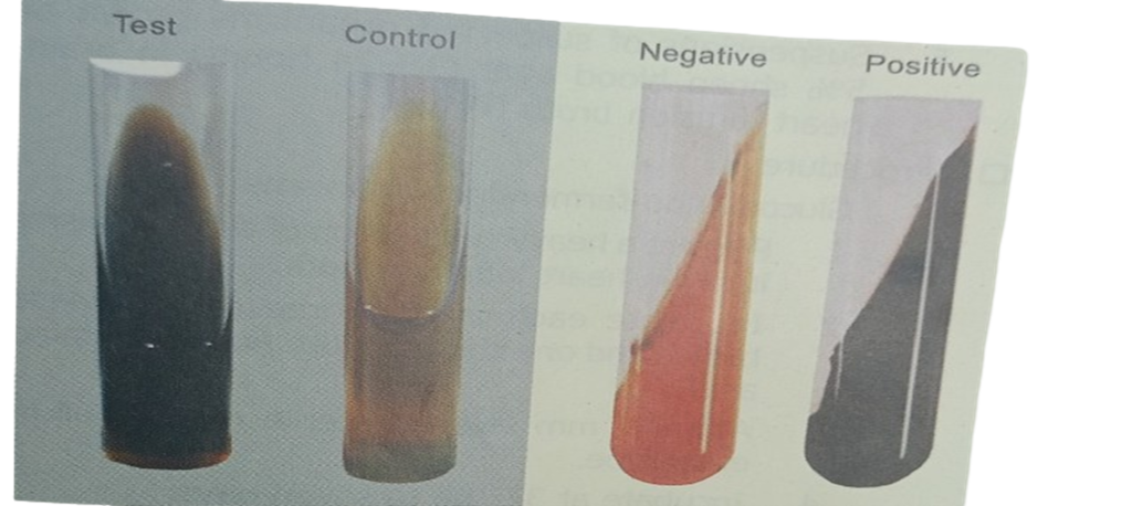
NITRATE REDUCTION TEST:- This test helps to differentiate bacteria that produce the enzyme nitrate reductase from the bacteria that do not produce the enzyme. This test is also helpful in differentiating Mycobacterium species. v      Principle:-       The test organisms are incubated in a broth containing nitrate. The nitrate reductase producing organisms reduce nitrate to nitrite, which is tested by adding sulfanilic acid reagent and a-naphthylamine. The formation of pink red compound indicates positive reaction.    v Requirements:-        1.      Nitrate broth         2.      Sulfanilic acid reagent                 a) Glacial acetic acid                        : 5.7 ml  b) Distilled water                              :  14.3 ml c) Sulfanilic acid                                :  0.16 g  3. alfa-naphthylamine  reagent a) Glacial acetic acid                        :  5.7 ml b) Distilled water                               : 14.3 ml c) Sulphonic acid                               :0.16 g v     Procedure:- 1.      Incubate the nitrate broth (sterile) with test organism. 2.      Incubate at 37°C for four hours 3.      Add one drop of sulfanilic acid reagent. 4.      Add one drop of a naphthylamine reagent. 5.      Mix well and observe the reaction   v Observations:-        Red color                            : Positive test         No red color                       :No reduction of nitrate   v Additional information:-      When nitrate is not detected it is necessary to test whether the organism has reduced the nitrate beyond nitrite to nitrogen gas or ammonia. Zinc dust (knife point), small amount is added, which will convert any nitrate to nitrite. In that case the observation is as follows:       Red color             : Negative test       No red color        : Positive test v    Quality control:- Use the following microorganisms to confirm the reliability of reagents:- Positive control        : E.coli Negative contro l      : Acinetobacter sp.

Microbiology, Uncategorized

INDOLE TEST FOR BACTERIA IDENTIFICATION

INDOLE TEST :- This test is important in the identification of enterobacteria such as E.coli, P. vulgaris, P. rettgeri, etc. v Principle:- The test organism is cultured in a medium containing tryptophan. The organisms break down tryptophan and indole is released. It is detected by the action of Kovac’s or Ehrlich reagent (formation of red colored compound. This test can also be performed by culturing the organism in tryptone water or peptone water containing tryptophan. Indole production is detected as described above). v   Requirements:- 1.      Motility indole urea (MIU) medium 2.      Kovac’s reagent strips (or Kovac’s reagent) Kovac’s Reagent v  Formula and preparation:- 1. p-dimethylaminobenzaldehyde            :        2.0 g 2. Isoamyl alcohol                                       :       30 ml 3. Concentrated hydrochloric acid           :       10 ml Dissolve ingredients 1 and 2 in 10 ml of concentrated hydrochloric acid and store in a brown bottle. v Procedure:- 1.      Inoculate MIU medium with test organism colonies. 2.   Incubate at 37°C by placing Kovac’s reagent strip in the neck of the MIU tube and 3 .       Look for reddening of the lower part of the test strip (or formation of red color of the reaction mixture). v Results:-  1. Reddening of strip                  : Positive test 2  No red color                              : Negative test v Quality control:- Use following microorganisms to confirm the reliability of reagents Positive control: Escherichia coli Negative control: Klebsiella aerogenes

Microbiology, Uncategorized

UREASE TEST FOR BACTERIA

Proteus strains are strong urease producers. Salmonellae and Shigellae do not produce urease. This test helps in differentiating enterobacteria. v    Principle:-  The test organisms are cultured in MIU medium (or in Christensen’s urea broth). If the strain produces urease, it acts on urea, and ammonium carbonate is formed with the release of ammonia. The medium becomes alkaline and the color of the medium changes to red pink. v  Requirements:- 1.      Motility Indole Urea (MIU) medium. 2.       Test tubes (15 x 125 mm) v  Procedure:- 1.      Inoculate MIU medium with acolony of the test organism. 2.      Incubate at 35°C overnight. 3.      Examine the medium by looking for a red pink color. v    Observations:- 1.     Red-pink medium       :  Positive test 2.     No red pink color        :  Negative test v  Quality control:-    Use the following microorganisms to confirm the reliability of reagents:-    Positive control     :           Klebsiella aerogenes    Negative control    :         E. coli

Microbiology

ESCULIN HYDROLYSIS TEST

v Principle:- This test is used to identify the bacteria (such as Klebsiella pneumonia) which are able to hydrolyze esculin (a glycoside). v Requirements:- 1.      Slant containing esculin 2.      24 hour broth culture  v Procedure:- 1.      Inoculate the medium with one drop of 24 hour 2.      broth culture 3.      Examine the slants v Observations:- 1.      Blackening of slant: Klebsiella pneumonia. (Positive test) 2.      No blackening of slant: Negative test When observed for fluorescence under Wood’s lamp, or positive test, loss of fluorescence is seen under Wood’s lamp. In the case of negative test there was no loss of flfluorescence. v Quality control:- 1.       Use following microorganisms to confirm the reliability of reagents: 2.       Positive control: Klebsiella pneumonia Negative test: Shigella flexneri

Microbiology

DECARBOXYLASE TESTS (MOELLER’S METHOD)

v Principle:- This test is used to detect the enzymatic ability of an organism  to decarboxylase (hydrolyse) an amino acid toform an amine. Hydrolysis of an amino acid results in an alkaline pH change leading to formation of pink color.  formation pink color. vRequirements:- 1.      Glucose non-fermentingorganisms 2.      Glucose fermenting organisms 3.      Decarboxylase broaths (lysine, arginine, ornithine) 4.      Sterile mineral oil 5 Suspensions of suspected organisms grown on 5% sheep blood agar (18-24 hours) in brain- heart infusion broth (BHIB). v Procedure:- A.    Glucose non-fermenting organisms 1.       Prepare a heavy suspension of the organisms in brain-heart infusion broth. 2.       Inoculate each of the three decarbxylase broths and one control broth  (without amino   acid). 3.       Add a 4 mm layer of sterile mineral oil in each tube. 4.       Incubate at 37° up to seven days. 5.       Observe color change.   B.       Glucose fermenting organisms 1.       Inoculate each of the decarboxylase tubes with 1 drop of brain-heart infusion broth   culture. 2.       Add 4 mm layer of sterile mineral oil in each tube. 3.       Incubate at 37° up to seven days. 4.       Observe color change. vObservations: By comparing with the color of the control tube:-   1   Positive:   Purple color (alkaline color change) Lysine:    Klebsiella pneumonia Arginine:     Enterobacteria cloacae Ornithine:    Enterobacteria cloacae  Negative:     No color change or yellow color                             (Due to fermentation of glucose in BHIB).   v  Usefollowing microorganisms to confirm the reliability of reagents: 1   Positive test:    Lysine:      Klebsiella pneumonia Arginine:   Enterobacter cloacae Ornithine:  Enterobacter cloacae Negative test:   Lysine:      Enterobacter cloaeae Arginine:    Klebsiella pneumonia Ornithine:   Klebsiella pneumonia v Note 1         Reference Book Text book of medical laboratory Technology by Dr. Praful B. Godkar 

Microbiology

OXIDASE TEST FOR BACTEIRA

This test is usedto help in the identification of the organisms which produce the enzyme oxidase. Examples:   Pseudomonas, Neisseria, Vibrio and Pasteurella species. v Principle:- A colony of the test organism is smeared on a filter paper piece, soaked with few drops of oxidase reagent. If the organisms are oxidase producing, the phenylenediamine in the reagent is oxidized to a deep purple color.  Requirements:- Oxidase reagent: 1.0 g/dl tetramethyl p- phenylenediamine dihydrochoride in distilled water (it should be prepared fresh if it appears blue in color.)          2  filter paper strips v Procedure:- 1.           Place a piece of filter paper a clean petri dish. 2.           Add 2 to 3 drop of freshly prepared oxidase reagent. 3.          Smear a colony of the organism on the filter paper by using aglass rod. v Observation:- 1.                Blue purple color : positive test 2.                No blue purple color : negativetest v   Quality control:- Use following microorganisms to confirm the reliability of reagents: Positive control: Pseudomonas aeruginosa Negative control: E. coli

Microbiology

HYDROGEN SULFIED PRODUCTION

This test is performed mainly to assist in the identification of Enterobacteria.      v Principle:-                 When sulfur-containing amino acids are decomposed by the enterobacteria, hydrogen sulfide is                           produced. It reacts with ferrous ions and the formation of ferric sulfide imparts a black color to the                   medium.     v Requirements:-            1.      TSI medium         v Procedure:- 1.      Stab the butt and streak the slant at 37°C 2.      Observe daily for seven days for blackening caused by H,S.     v    Results:-                   1.           Blackening: Positive test.                      2.           No blackening: Negative test,No HS production.                v Quality control:- Use following microorganisms to confirm the reliability of reagents: Positive control: Staphylococcus aureus Negative control: Escherichia coli

Microbiology

CITRATE UTILIZATION TEST

This test is performed in the identification of Enterobacteria:-     v Principle:- The test organism is cultured in a medium containing sodium citrate, an ammonium salt and a bromothymol blue indicator. The organisms use citrate (the only source of carbon) and ammonia (the only source of nitrogen). The citrate utilization is followed by an alkaline reaction (change of the color from light green to blue) and growth in the medium is indicated by the appearance of turbidity.     v Requirements           Test tube (15 x 125 mm),                 Simmon’s citrate medium,               Formula and preparation  1.      Potassium dihydrogen phosphate                    : 1.0 g 2.      Sodium ammonium phosphate                          : 1.5 g 3.      Magnesium sulfate                                               : 0.2 g 4.      Sodium citrate                                                        : 2.5 g 5.      Bromothymol blue                                                 :0.016 g 6.      Agar                                                                          :15.0g 7.      Distilled water                                                           1000ml The ingredients 1 to 6 are dissolved in about 900 ml of distilled water and after diluting to one liter, pH of this medium is adjusted to 6.7-6.9.     v Procedure            1.      Inoculate 3-4 ml of sterile Simmon’s citrate medium with a broth culture of test organisms.               2.      Incubate at 35°C up to 4 days.     v Results           1.       Turbidity and blue color: Positive test.              2.     No growth (no turbidity and persistence of original color): Negative test.     v Quality control            Use following microorganisms to confirm the reliability of reagents:              Positive control: Klebsiella pneumonia  Negative control: Escherichia coli

Microbiology

DITECTION OF COAGULASE ENZYME TEST

COAGULASE TEST:- This test is used to differentiate Staphylococcus aureus from S. epidermidis and S. saprophyticus –     v    Principle:-           Staphylococcus aureus produces the enzyme coagulase which causes plasma to clot                          converting soluble  fibrinogen to insoluble fibrin.     v  Requirements:- 1.      Oxalate or citrated plasma 2.      Glass slides 3.      Normal saline     v Procedure (Slide Method):- 1.      Place a drop of physiological saline on each end of a slide. 2.      Make thick suspensions of the organisms in each drop. 3.      Add a drop of plasma to one of the suspensions. Mix gently. Look for clumping of the organisms within 10 seconds.      v Results:-             1.      Clumping within 10 seconds: Staphylococcus aureus               2.      No clumping within 10 seconds: No production of coagulase  v Tube test Method:- 1.      Prepare 1.0 ml of saline suspension of the organism 2.      Add 1.0 ml of plasma 3.      Incubate at 37°C for 24 hours. 4.      Observe from clot formation. The formation of a clot indicates the presence of Staphylococcus aureus organism.

Microbiology

CATALASE TEST

SUMMARY:- This test is used to differentiate catalase-producing bacteria such, as Staphylococci from non-catalase-producing bacteria such as Streptococci.     v   Principle:-           Catalase produced by the organisms acts on hydrogen peroxide to produce water and oxygen (It is                     indicated by bubbles).     v  Requirements:-           1.      3% (v/v) Hydrogen peroxide              2.       Test tubes (15 x 125 mm)     v Procedure:- 1.       Pour 2 to 3 ml of hydrogen peroxide solution into a test tube. 2.       Immerse the growth of organisms in the test tube solution by using a sterile wooden stick (or a sterile glass rod). 3.       Look for immediate bubbling.     v   Observations:-           1.      Appearance of bubbles: Presence of catalase-producing organisms.              2.      No formation of bubbles: Presence of non-catalase-producing organisms.     v   Precautions:-           1.      The culture should not be for more than 24 hours.              2.      This test should be performed from a blood-free culture medium (nutrient agar) since catalase is                      also  present in red blood cells              v Quality control:- Use the following microorganisms to confirm the reliability of reagents: 1.      Positive control: Staphylococcus species 2.      Negative control: Streptococcus species

Scroll to Top