Microbiology

Microbiology explores the world of microorganisms such as bacteria, viruses, fungi, and parasites. It is vital for understanding infections, disease transmission, sterilization, and laboratory identification of pathogens.

Microbiology

MOTILITY TESTING OF MICROORGANISUM

Motility testing of the microorganism or hanging drop preparation:- Requirements:- 1.       Hollow ground (depression) or cavity slide 2.       Coverslip 3.       Petroleum jelly 4.       Two known cultures (one containing motile and other non-motile organism) 5.       Microscope Procedure– 1.      Apply petroleum jelly at the four corners of the coverslip. 2.       Place aseptically loopful of culture at the middle of the coverslip. 3.       Take the cavity slide and invert the concave portion over the drop. 4.       Invert the whole preparation so that the coverslip is on the top. 5.      Examine the suspension under low power lens first. Cut down the light by adjusting the diaphragm of the condenser and then swing the objectives to the high dry lens, 6.      Observe for motility (it can be detected by the presence of directional motion). Note– 1.       Look at one organism for about 1 minute and see if it moves out of its original position. 2.       Jumping or dancing organisms are not necessarily true motiles, but these movements may be due to Brownian movement. 3.       Record the results of the observations by indicating “motile” or “non-motile” organism. After finishing the observations, place the cavity slide in disinfectant solution.

Microbiology

Gram staining Theories

Gram stain:- Very commonly used differential staining is Gram staining, used to differentiate gram-positive & gram-negative bacteria. Developed by Hans Christian Gram in 1884. Gram’s Staining Principle:- 1.   Bacteria having cell walls with a thick layer of peptidoglycan will resist decolorization of primary stain and appear violet or purple. 2.    Bacteria having a thin peptidoglycan layer with lesser cross-linkage lose primary stain during decolorizing and gain counter stain appearing pink orred.  3.    In an aqueous solution of crystal violet dye, their molecules dissociate into CV+ and Cl– ions. These ions easily penetrate the cell wall components of both positive and negative bacteria. 4.  When Gram’s Iodine is added as mordant, the iodine (I) interacts with CV+ion and forms CV-I complex within cytoplasm and cell membrane and cell wall layers. 5.When decolorizing solution (ethanol or a mixture of ethanol and acetone) is added it interacts with lipids in the cell wall. 6.  The outer membrane of the Gram-Negative bacterial cell wall is dissolved exposing the peptidoglycan layer 7.     The peptidoglycan layer is thin with less cross-linking in the Gram-Negative cell wall, hence becoming leaky. This causes cells to lose most of the CVI complexes. 8.Whereas in Gram-Positive bacteria, there is no outer membrane, and the peptidoglycan layer is also thick with higher cross-linkage. 9.So, the decolorizing solution dehydrates the peptidoglycan layer trapping all the CVI complexes inside the cell wall and bacteria retain the purple orviolet color of crystal violet. 10.When counterstain, positively charged safranin, is added, it interacts with the free negatively charged components in Gram-Negative cell wall and membrane and bacteria becomes pink/red. 11.Whereas, there is no space to enter inside the dehydrated Gram-Positive cell wall due to CVI complex and dehydration. Hence, safranin can’t stain themred or pink and Gram-Positive bacteria reveal the purple or violet color. Gram’s Staining Protocol:- •       Flood crystal violet solution over fixed smear •       After 30 – 60 seconds, pour off the Crystal Violet solution and rinse with gentle running water. •       Flood the Gram’s Iodine solution over the smear •       Leave the iodine solution for 30 – 60 seconds and pour off the excess iodine and rinse with gentle running water •       Shake off the excess water over the smear •       Decolorize the smear by passing the decolorizing solution till the solution runs down in clear form. Alternatively, add a few drops of decolorizing solution and shake gently and rinse with distilled water after 5 seconds. •       Rinse with distilled water to wash decolorizer •       Shake off the excess water over the smear •       Pour counter stain over the smear •       Leave for 30 – 60 seconds and wash with gentle running water •       Air-dry or blow-dry the smear.

Microbiology

BACTERIAL DISEASES

PULMONARY TUBERCULOSIS :- Pathogen:-      Mycobacterium tuberculae Incubation Period:-      2-10 weeks Symptoms:-Coughing; chest pain and bloody sputum with tuberculin. Epidemiology:-Airborne & Droplet infection Therapy/ Prophylaxis:-Streptomycin, para-amino salicylic acid, rifampicin etc./ BCG vaccine Isolation, Health education DIPHTHERIA:- Pathogen:- Corynebacterium diphtheriae Incubation Period:-     2-6 days Symptoms:-Inflammation of mucosa of nasal chamber, throat etc. respiratory tract blocked. Epidemiology:-Airborne & Droplet infection Therapy/ Prophylaxis:-Diphtheria antitoxins, Penicillin, Erythromycin/ DPT vaccine CHOLERA:- Pathogen:-   Vibrio cholerae Incubation Period:-6 hours to 2 – 3 days Symptoms:-Acute diarrhoea & dehydration. Epidemiology:-Direct & oral (with contaminated food & water) Therapy/Prophylaxis:-Oral rehydration therapy & tetracycline/ Sanitation, boiling of water & cholera vaccine Leprosy:- Pathogen:-Mycobacterium leprae Incubation Period:-  2-5 years Symptoms:-Skin hypopigmentation, nodulated skin, deformity of fingers & toes. Lepromin in skin tests. Epidemiology:-lowest infectious & contagious Therapy/Prophylaxis:-Dapsone, rifampicin, Clofazimine./ Isolation Pertussis(Whooping Cough):- Pathogen:-Bordetella pertussis Incubation period:- 7-14 days Symptoms:-Whoops during inspiration Epidemiology:-Contagious & Droplet infection Therapy/Prophylaxis:-Erythromycin / DPT vaccine Tetanus(Lock Jaw):- Pathogen:-Clostridium tetani Incubation Period:- 3-21 days Symptoms:-Degeneration of motor neurons, rigid jaw muscles, spasm and paralysis Epidemiology:-Through injury Therapy/Prophylaxis:-Tetanus- antitoxins. / ATS and DPT vaccines. Gonorrhoea:- Pathogen:-          Neisseria gonorrhoeae Incubation Period:-   2-10 days Symptoms:-   Inflammation of urinogenital tract. Epidemiology:-   Sexual transmission Therapy/Prophylaxis:-Penicillin & Ampicillin / Avoid prostitution Syphilis:- Pathogen:-Treponema pallidum Incubation period:-3 weeks Symptoms:-Inflammation of urinogenital tract. Epidemiology:-Sexual transmission Therapy/Prophylaxis:-Tetracycline & penicillin. / Avoid prostitution Viral Disease:- Chickenpox(Varicella): Pathogen – Herpes-zoster virus (DNA- virus) Epidemiology – Contagious & Formite borne Incubation Period – 12-20 days Symptoms – Dark red coloured rash or pox changing into vesicles, crusts and falling. Prophylaxis – Now vaccine available, isolation. Therapy – Zoster Immunoglobulins (ZIG). 2. Smallpox(Variolla):- Pathogen – Variola-virus (DNA-Virus) Epidemiology – Contagious & Droplet infection Incubation Period – 12-days Symptoms – Appearance of rash changing into pustules, scabs and falling. Prophylaxis – Smallpox vaccine. Therapy – No case reported after 1978. 3.Poliomyelitis:- Pathogen – Polio-virus (RNA-virus) Epidemiology – Direct & oral Incubation Period – 7-14 days Symptoms – Damages motor neurons causing stiffness of neck, convulsion, paralysis of generally legs. Prophylaxis – ‘Salk’ vaccine and Oral Polio vaccine. Therapy – Physiotherapy. 4. Measles(Rubeolla Disease):- Pathogen – Rubeolla-virus (RNA-virus) Epidemiology – Contagious & Droplet infection Incubation Period – 10 days Symptoms – Rubeolla (skin eruptions), coughing, sneezing etc. Prophylaxis – Edmonston- B-vaccine, isolation Therapy – Antibiotics & sulpha drugs. 5. Mumps:- Pathogen – Mumps-virus (RNA-virus) Epidemiology – Contagious & Droplet infection Incubation Period – 12-26 days Symptoms – Painful enlargement of parotid salivary glands. Prophylaxis – Mumps- vaccine isolation Therapy – Antibiotics. 6. Rabies(Hydrophobia) Pathogen – Rabies-virus (RNA-virus) Epidemiology – Indirect & inoculative (vectors are rabid animals monkeys, cats, dogs) Incubation Period – 10 days to 1- 3 months Symptoms – Spasm of throat & chest muscles, fears from water, paralysis and death. Prophylaxis – Immunization of dogs. Therapy – Pasteur- treatment (14 vaccines in stomach). 7.Trachoma:- Pathogen – Chlamydia trachomatis Epidemiology – Contagious, formite-borne and flies (vectors) Incubation Period – 5-12 days Symptoms – Inflammation of conjunctiva & cornea leading to blindness. Prophylaxis – Isolation Therapy – Tetracycline & sulfonamide. 8.Influenza(Flu):- Pathogen – Myxovirus influenzae (RNA-virus) Epidemiology – Air borne and pandemic Incubation Period – 24-48 Hours Lasts for 4-5 days Symptoms – Bronchitis, sneezing bronchopneumonia, leucopenia, coughing, etc. Prophylaxis – Isolation. Therapy – Antibiotic therapy. 9.Hepatitis(Epidemic Jaundice):- Pathogen – Hepatitis-B virus Epidemiology – Direct & oral (with food and water) Incubation Period – 20-35 days Symptoms – Damage to liver cells releasing bilirubin which causes jaundice. Prophylaxis – Proper sanitation proper coverage of food, water, milk etc. use of chlorinated or boiled water, etc. Therapy – Hepatitis-B vaccine.

Microbiology

MICROBIOLOGY AND SEROLOGY PRACTICAL INDEX

MICROBIOLOGY INDEX 1.      Safety measures in the Microbiology Laboratory working area. 2.      Isolation of bacteria 3.      Culture media a.     Introduction and preparation technique of     Culture Media b.    Type of Culture Media       Nutrient Agar       MacConkey Agar        Blood Agar         EMB Agar         XLD Agar          Deoxycholate Citrate Agar          Lowenstein Jensen Acid Medium 4.      Isolation of Bacteria       a.  Inoculation Technique           Streaking Method,     Pouring Method     Spreading Method       Lawn Method b. Incubation of micro-organisms on or in Culture media c.  Study of Bacterial Colony morphology d.   Biochemical identification of microbiology   i.     Catalase Test ii.     Coagulase Test iii.     Oxidase Test iv.     Urease Test v.     Indole Test vi.     Bile Solubility test vii.    Cetrimide test viii.  Decarboxylase test.       e.     Slide preparation of various Specimens f.     Staining Technique  i.     Simple Stain  ii.   Gram’s Stain  iii. AFB or ZN Stain   iv.  Albert Stain   v.     Negative Stain 5.       Cultivation of Fungi 6.       Motility Testing of micro-organisms 7.       Anti-bacterial sensitivity testing 8.       Preparation of VDRL buffer and Antigen emulsion Serology practical index 1.       VDRL Agglutination test for syphilis by Quantitative and Qualitative method 2.       Rapid Plasma Reagin (RPR) test for syphilis 3.       Widal test by Qualitative and quantitative method. 4.       Ring Test 5.       Nepier Aldehyde Test 6.       ASO test by Quantitative and Qualitative 7.       Rheumatoid Arthritis Test (RA Test) 8.       C-Reactive Protein Test (CRP)   9.       Immunologic Pregnancy Test

Microbiology

STERILIZATION

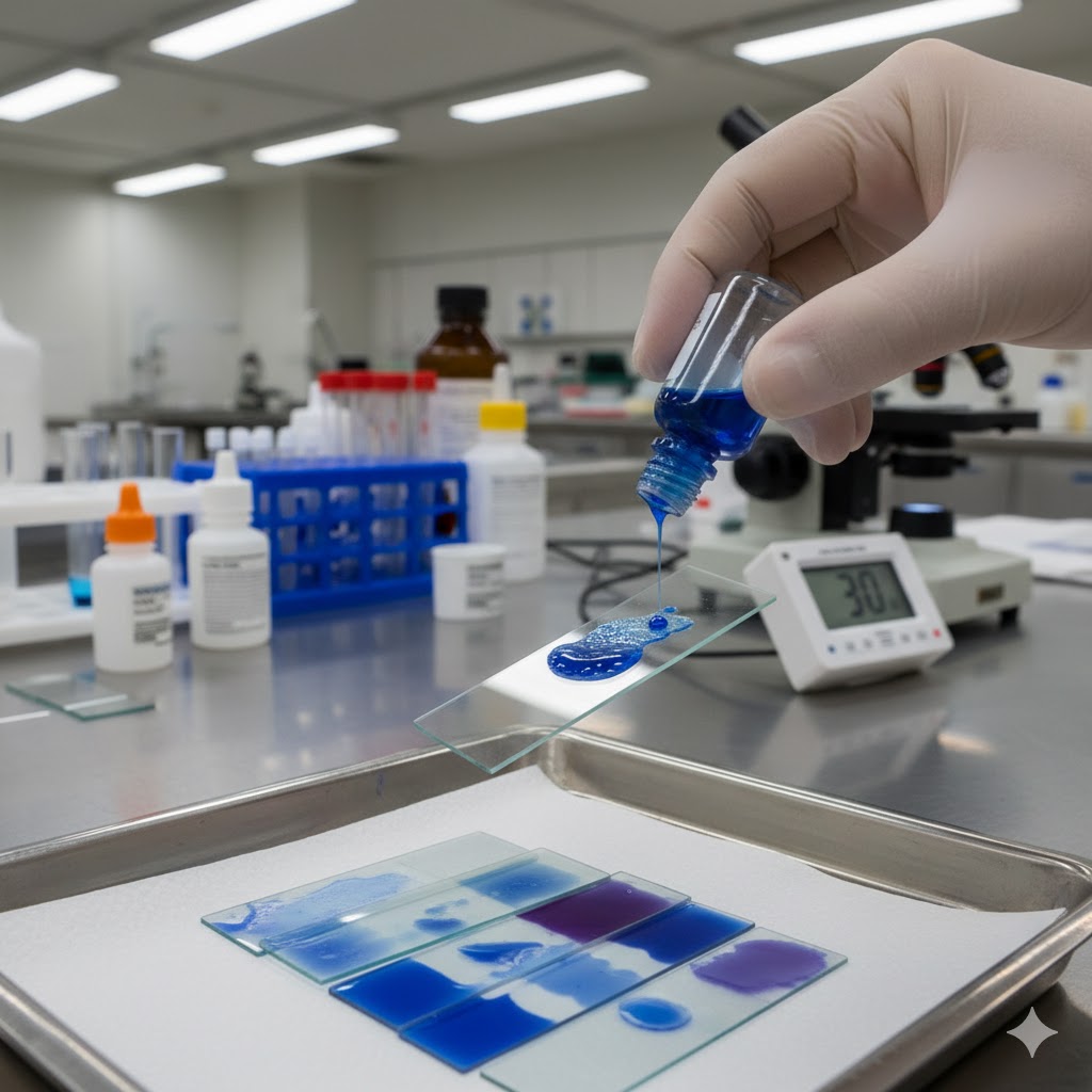
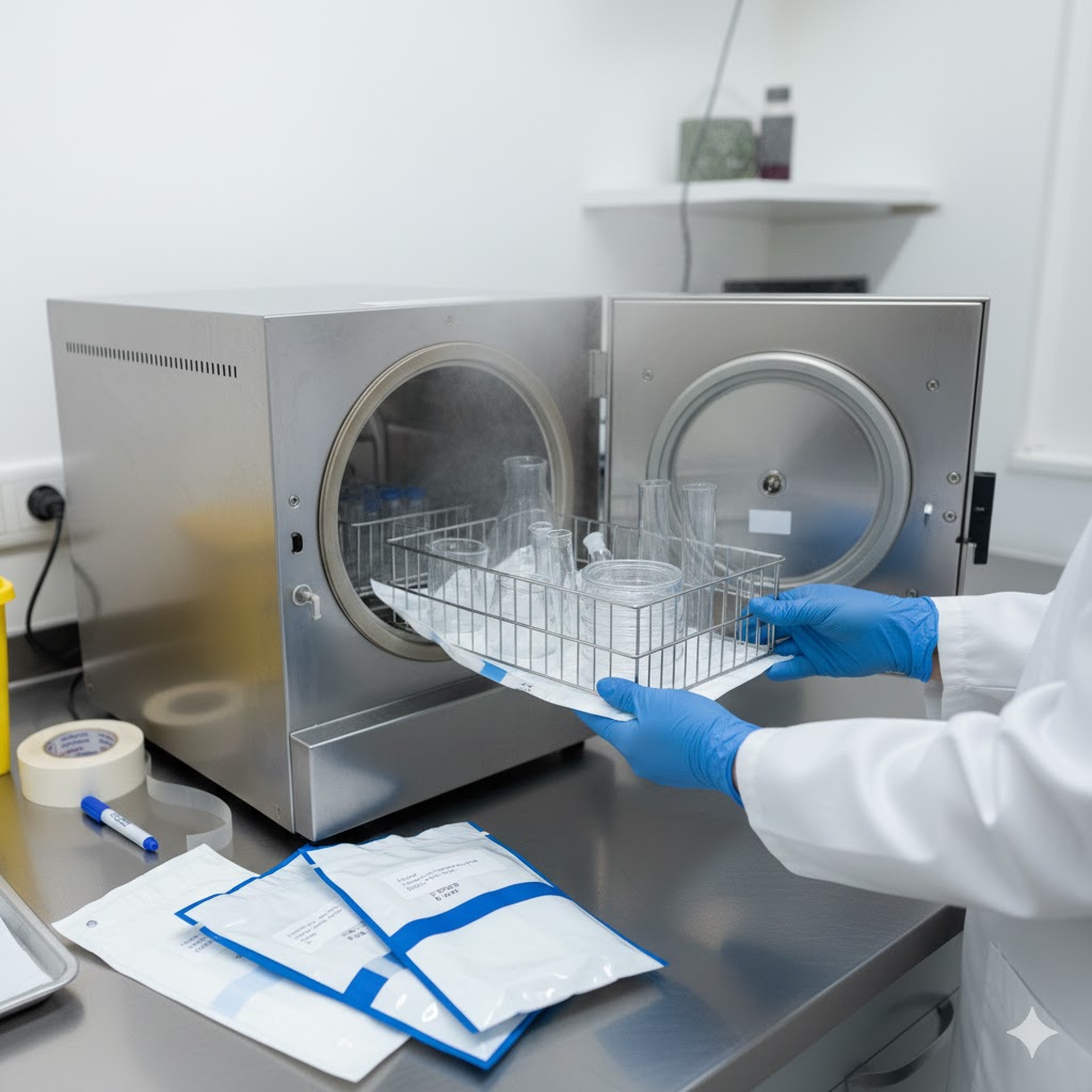
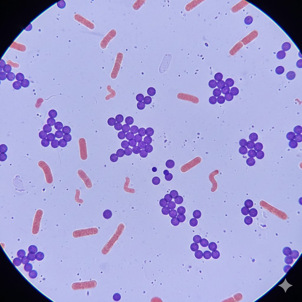
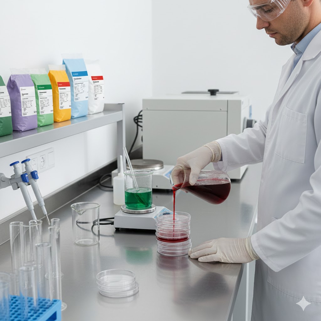
STERILIZATION:- Sterilization (or sterilisation) refers to any process that eliminates, removes, kills, or deactivates all forms of life and other biological agents.(suchas:- fungi, bacteria, viruses, spore forms, prions, unicellular eukaryotic organisms such as Plasmodium, etc) present in a specified region, such as a surface, a volume of fluid, medication, or in a compound such as biological culture media. Sterilization can be achieved through various means, including: heat, chemicals, irradiation, high pressure, and filtration. Sterilization is distinct from disinfection, sanitization, and pasteurization, in that sterilization kills, deactivates, or eliminates all forms of life and other biological agents which are present.   Sterilization are classified asfollowing:- physical methods of seterilization (i)Sun-Light:- Ultraviolet rays present in the sun-light are responsible for spontaneous sterilization in natural conditions. In tropical countries the sun light is more effective in killing bacteria due to combination of ultraviolet rays and heat. By killing bacteria in suspended water, sunlight provides natural method of disinfection of water bodies such as tanks and lakes. Those articles which cannot withstand high temperature can still be sterilised at lower temperature by prolonging theduration of exposure. (ii)Heat:- It is considered to be the most reliable method of sterilization of articles—that withstand heat. There are two methods of the sterilization: dry heat and moist heat. (A) Dry Heat: It acts by protein denaturation and oxidative damage. Sterilization by dry heat is as follows:-  (a) Red Heat: Articles such as bacteriological loops, straight wires, tips of forceps and searing spatulas are sterilised by holding them in a Bunsen flame, till they become hot red. (b) Flaming: Articles are passed over a Bunsen flame but not heating it to redness. (c) Incineration: Contaminated, materials are destroyed by burning them in incinerator. (d) Hot Air Oven: Articles are exposed to high temperature (160°C) for duration of one hour in an electrically heated oven (method was introduced by Louis Pasteur). (B) Moist Heat: It acts by coagulation and denaturation of proteins. (i)At Temperature below 100°C:   Pasteurization: This process was originally employed by Louis Pasteur. Currently this procedure is employed in food and dairy industry. There are two methods of pasteurization, the holder method (heated at 63oC for 30 minutes) and flash method (heated at 72oC for 15 seconds) followed by quickly cooling to 13oC. Other pasteurization methods include Ultra-High Temperature (UHT), 140oC for 15 sec and 149oC for 0.5 sec. This method is suitable to destroy most milk borne pathogens like Salmonella, Mycobacterium, Streptococci, Staphylococci and Brucella, however Coxiella may survive pasteurization. Efficacy is tested by phosphatase test and methylene blue test. Vaccine bath:- The contaminating bacteria in a vaccine preparation can be inactivated by heating in a water bath at 60oC for one hour. Only vegetative bacteria are killed and spores survive.  Serum bath:- The contaminating bacteria in a serum preparation can be inactivated by heating in a water bath at 56oC for one hour on several successive days.Proteins in the serum will coagulate at higher temperature. Only vegetative bacteria are killed and spores survive. (ii) At Temperature 100°C:- (a)Boiling:- Boiling water (100C°) kills most vegetative bacteria. (b) Steam at 100°C:- Passing the steam at 100°C over articles kills bacteria. Sugars and gelatin in medium may get decomposed by autoclaving. So, these can be sterilised by exposing them to free steaming for 20 minutes for three successive days. This process is known as tyndallisation (after John Tyndall). (iii)At Temperature above   100°C:- (a) Autoclave:- Sterilization can be effectively achieved at a temperature above 100°C using an autoclave. Structure of Autoclave:- A simple autoclave has vertical or horizontal cylindrical body with a heating element, a per-forted tray to keep the articles, a lid that can be fastened by screw clamps, a pressure gauge, a safety valve and a discharge tap (Fig. 1). The lid is closed but the discharge tap is kept open and the water is heated. As the water starts boiling the steam drives air out of the discharge tap, when all the air is displaced and steam starts appearing through the discharge tap, the tap is closed. The pressure inside is allowed to rise up to 15 lbs. per square inch. At this pressure the articles are heated for 15 minutes, after which the heating is stopped and the autoclave is allowed to cool. Once the pressure gauge shows the pressure equal to  atmospheric pressure, the discharged tap is opened to let the air in. The lidis opened and articles are removed. Culture media, dressing, certain equipment’s can be sterilised by autoclave. (iii) Sonic and Ultrasonic Vibrations- Sound waves of frequency 720,000 cycle/second kills bacteria and some viruses exposing for one hour. (iv) Radiation:- Two types of rays are used for sterilization: Non-ionizing and ionizing. (a) Non-Ionizing Rays:- These are low-energy rays with poor penetrative power, e.g., U.V. rays (wavelength 200-280 nms, effective 260 nm). (b) Ionizing Rays:- These are high-energy rays with good penetration power. These are of two types:- Particulate and electromagnetic.Electron beams are particulate while gamma rays are electromagnetic in nature. High speed electrons are produced by a linear accelerator from a heated cathode.Electromagnetic rays such as gamma rays emanate from nuclear disintegration of certain radioactive isotopes (Co60, Cs137). A degree of 2.5 megabrands of electromagnetic rays kills all bacteria, fungi, virus and spores. In some parts of Europe, fruits and vegetable are irradiated to increase their shelf life up to 500 percent. Since radiation does not generate heat, it is called Cold sterilization.  Chemical Methods of Sterilization: Chemicals destroy pathogenic bacteria from inanimatesurfaces and are all also called disinfectants. Liquid:- Alcohols:- E.g., Ethyl alcohol, Isopropyl alcohol and methyl alcohol.  (A 70% solution kills bacteria). Aldehydeles: E.g.,Fomaldehyele,Gluteraldehydele(40% formaldehyde is used for surface disinfection). Phenol:- E.g., 50% phenol, 1-5%cresol, 5% lysol, chloroxylenol (Dettol). Halogens: E.g.,chlorine compounds (chlorine bleach, hydrochloride) and iodine compounds (tincture, iodine, iodophores). Tincture ofiodine (2% iodine in 70% alcohol) is antiseptic. Heavy metals:- E.g., Mercuric chloride, silver nitrate, copper sulfate, organic mercuric salts. Surface active agents: e.g., soaps or detergents. Dyes:- Acridin dyes e.g., acriflavin and aminacrine are bactericidal (interact with bacterial nucleic acids). Gaseous: E.g.Ethylene oxide, formaldehyde gas, highly effective, killing of spores. Physiochemical Methods of Sterilization: A physiochemical method adapts both physical and

Microbiology

GRAM STAIN

GRAM STAIN :- Objective :- To differentiate bacteria into Gram-positive and Gram-negative based on the ability of their cell wall to retain the primary stain (crystal violet) after decolorization. Principle Gram-positive bacteria: Thick peptidoglycan layer → retain crystal violet–iodine complex → appear purple. Gram-negative bacteria: Thin peptidoglycan, high lipid content → lose crystal violet on decolorization → take up safranin → appear pink/red. Materials Required Clean glass slides Inoculating loop / needle Bunsen burner Staining rack Wash bottle with water Blotting paper Reagents Crystal Violet (Primary stain) Gram’s Iodine (Mordant) Decolorizer (Acetone–alcohol or 95% ethanol) Safranin (Counterstain) Procedure 1. Preparation of Smear Clean the slide and label it. Place a small drop of water on the slide. Pick a small amount of culture and spread to form a thin smear. Air dry completely. Heat fix by passing the slide over flame 2–3 times (do not overheat). ·         2. Gram Staining Steps Step Reagent Time   1 Crystal Violet 1 minute 2 Wash gently — 3 Gram’s Iodine 1 minute 4 Wash gently — 5 Decolorizer (Alcohol/Acetone) Few seconds (5–15 sec) 6 Wash immediately — 7 Safranin 30–60 seconds 8 Final wash — 9 Blot dry — Microscopic Examination Observe under oil immersion (100x) objective. Findings Gram-positive → Purple / violet Gram-negative → Pink / red Note shape: cocci, bacilli, spirilla, clusters, chains, pairs.

Microbiology

CULTURE MEDIA INTRODUCTION &PREPARATION

INTRODUCTIONOF CULTURE MEDIA Most bacteria can be cultured artificially on culture media containing required nutrients, pH and osmotic pressure. The microorganisms grow in an atmosphere and temperature most suited to their metabolic reactions. The pathogens are isolated in pure culture so that they can be identified and tested for their sensitivity to antimicrobials. The specimens are cultured in known volumes, and the number of bacterial colonies appearing after incubation can be counted. Operation room requirements and blood from blood bank are frequently checked for sterility by using pure culture methods. Vaccines and antitoxins require the growing of bacteria under controlled conditions. Stock cultures are also useful for the teaching institutes for practical training purposes. COMPOSITION OF CULTURE MEDIA The basic ingredients, which are common to the many of the frequently used media are as follows: 1.  Water: It allows fluids to enter and leave cells more readily and to enhance the chemical reactions. 2.Sodium chloride: Presence of sodium chloride maintains the isotonicity  of bacterial cells. 3.Peptones: It is a source of readily available nitrogen. 4.Buffers: They maintain a constant pH in culture media. 5.Indicators:  In culture media the indicators are useful in the detection of acid or alkali production by microorganisms. Indicators such as phenol red, methyl red and Bromocresol purple are used to adjust pH of the culture media. 6.  Solidifying agents: Agar, gelatin, egg yolks and serum are used as solidifying agents of the culture media. 7.Selective agents: These are special chemicals introduced into the culture media for inhibiting some types of bacteria, while allowing other bacteria to grow. Examples: (a) Crystal violet inhibits Staphylococci but not tubercle bacilli (b) 6.5% (w/ v) sodium chloride is inhibitory to most Streptococci but not S. faecalis. 8.   Additive for enrichment: The substances such as sheep blood, horse blood, rabbit serum or calf’s ground hearts allow fastidious organisms to grow since these organisms, may not survive in ordinary culture media. 9.Reducing substances: The substances such as thioglycollate are used to remove free oxygen from the medium for the growth of anaerobic bacteria. THE DIFFERENT TYPES OF CULTURE MEDIA Basic Media These support the growth of microorganisms that do not have special nutritional requirements. They are often used (a) To maintain stock cultures of control strains of bacteria and (b) For subculturing pathogens from selective media prior to performing biochemical and serological dentification tests. Examples: (1) Nutrient agar (2) Nutrient broth. Enriched Media These are enriched with (a) Whole blood (b) Lysed blood (c) Serum (d) Extra peptones and (e) Vitamins to support the growth of particular pathogens such as Hemophilus influenzae, Neisseria and Streptococcus species. Examples: (I) Blood agar (II) Tryptone soya media. Selective Media These media contain substances that accelerate the growth of required pathogens only and prevent or slow down the growth of other microorganisms. Example: XLD agar: It is used for the growth of Salmonellae and Shigellae. The bile salts present in this media inhibit the growth of many fecal commensals Differential (Indicator) Media These contain indicators, dyes or other substances which help to differentiate microorganisms. Example: TCBS agar contains the indicator bromothymol blue which differentiates sucrose fermenting from non-sucrose fermenting vibrio species. Transport Media When specimens are not cultured soon after collection, to prevent overgrowth and also to ensure survival of pathogens, transport media are used. These are mainly used to transport microbiological specimens from health centers to the district pathological laboratories. Example: (1) Amies transport medium (2) Cary Blair medium. DIFFERENT FORMS OF CULTURE MEDIA:- 1.      Solid Culture Media This is used in petri dishes and in test tubes (slope cultures) and prepared by adding Solidifying agent  (1.0 – 1.5%) (w/v). Microorganisms grow on this and form colonies after multiplication. This helps to identify the organism. 2.    Semisolid Culture Media This is prepared by adding Solidifying agent (0.4-0.5%) (w/v) to a fluid medium. These are used mainly as transport media and for the testing of motility of the organisms. 3.   Fluid Culture Media These media are mainly used as biochemical testing media, blood culture media or the enrichment media. PREPARATION OF CULTURE MEDIA Most culture media are available commercially in readymade dehydrated form. It is less costly to use readymade media, since the ingredients are often required in small amounts but available in large quantities if purchased. Some of the chemicals are also difficult to obtain. To ensure good performance and reproducibility in the results the following must be  performed correctly- 1.     Weighing and dissolving of the ingredients 2.     Addition of heat sensitive material 3.     pH testing 4.     Dispensing and sterilization 5.     Sterility testing and quality control 6. Storage Note 1.      The heat sensitive ingredients such as blood or serum should be brought to room temperature and added when the medium has cooled to about 50°C. 2.      A fluid medium should be tested for accurate pH by using a narrow, range pH paper. 3.      For sterilizing culture media, it is necessary to use manufacturer’s instructions. The commonly used methods for sterilization are- a) Autoclaving (b) Steaming at 100°C and (c) Filtration. It is necessary to use correct temperature and correct length of time. Precautions:- 1.  Autoclaving is used to sterilize most agar and fluid media. 2. Steaming at 100°C: Media such as Cary Blair transport medium contain ingredients that would break down above 100°C. Steaming can be performed in an autoclave with a loose lid. 3.    Filtration: Serum and solutions containing carbohydrates, urea, etc. are heat sensitive and hence cannot be autoclaved. Hence, the media containing such substances are filtered to remove bacteria. 4. Sterility testing: Media in tubes and bottles: Incubate the entire batch at 37°C overnight. Contamination is indicated by appearance of turbidity in a fluid medium and growth on a solid medium. 5.  Control of media: Appropriate control species are used to inoculate slants or plates of the medium (quarter part). After overnight incubation, the cultures are examined for (a) Degree of growth (b) Size of colonies and (c) Other characteristics. 6.Storage of culture media: Dehydrated culture media and dry ingredients (agars, peptones, bile salts, etc.) can be stored at room temperature (25°C ± 5°C) in a cool and dry place, away from

Microbiology, Uncategorized

INTRODUCTION OF MICROBIOLOGY

“Microbiology Worlds”  Microbiology is the scientific study of microorganisms, which are microscopic living organisms that include bacteria, viruses, fungi, and protozoa. These microorganisms play a crucial role in various biological processes and have a significant impact on human health, agriculture, industry, and the environment. French chemist and Microbiologist Louis Pasteur (1822-1895) coined the term “microbiology”. He used the term to describe his work with organisms at the microscopic level. The field of microbiology has a rich history that spans several centuries, with important discoveries and developments contributing to our understanding of the microbial world. Historical Background of Microbiology:- 1.0Aristotle (384-322 BC):-  The concept of spontaneous generation was proposed by various ancient civilizations and philosophers, including the ancient Egyptians and Greeks. One of the most notable proponents of spontaneous generation in ancient times was the Greek philosopher Aristotle. He proposed the idea that certain animals, insects, and even mice could arise spontaneously from decaying organic matter. In the Middle Ages, spontaneous generation was further supported by prominent figures such as Avicenna and Albertus Magnus. 2.0 Anton van Leeuwenhoek (1632 – 1723):- Anton van Leeuwenhoek made significant contributions to the field of microbiology, earning him the title of the  “father of microbiology & Protozoology.” Here are some of his key contributions: 1.  Discovery of Microorganisms:  In the 17th century, Leeuwenhoek designed powerful single-lens microscopes, enabling him to observe tiny organisms that were previously invisible to the human eye. He was the first to describe and document various microorganisms, including bacteria, protozoa, and other microscopic life forms.  In 1676 Observations of bacteria, which he called “animalcules,” provided crucial evidence against the theory of spontaneous generation. 2..  Microscopic Studies of Biological Samples: Leeuwenhoek examined a wide range of biological samples, including water, dental plaque, and even his own feces. 3. Advancements in Microscopy: Leeuwenhoek’s innovative improvements to microscope design, including using high-quality lenses and precise grinding techniques, significantly enhanced the magnification and clarity of his observations.  4..       The term “microbe” was first used by Sedillot in 1878. The word comes from the Greek words “mikros” meaning “small” and “bios” meaning “life”. It literally means “small life” or “microscopic life”. 3.0 Francesco Redi (1626-1697):- Francesco Redi was an Italian physician, naturalist, and poet who made significant contributions to the field of microbiology, particularly in the context of spontaneous generation, which was the widely held belief at the time, that living organisms could arise from non-living matter. Redi conducted a series of experiments that challenged this idea, providing evidence against spontaneous generation Francesco Redi experiments:- In 1668, Redi conducted a famous experiment to test the hypothesis that maggots (the larvae of flies) spontaneously generated from decaying meat. He set up three groups of jars, each containing decaying meat. One group was left open, allowing flies to access the meat and lay eggs, another group was covered with gauze, preventing flies from touching the meat but allowing air to pass, and the third group was completely sealed. Redi observed that maggots only appeared in the open jars, where flies could access the meat, and not in the covered or sealed jars. This experiment provided strong evidence against spontaneous generation, demonstrating that maggots only appeared when flies were able to lay their eggs on the decaying meat. 4.0 John Needham (1713 – 1781): In 1745, Needham conducted experiments where he heated nutrient broths and then sealed them in flasks. Afterward, he observed the growth of microorganisms in the sealed flasks and concluded that life could arise spontaneously from non-living matter. 5.0 Lazzaro Spallanzani (1729 – 1799):- Lazzaro Spallanzani Disproving Spontaneous Generation Theory and support Biogenesis Theory. In 1765, Lazzaro Spallanzani conducted experiments where he boiled nutrient-rich broth in sealed containers, effectively sterilizing the broth. His experiments showed that no microorganisms grew in the sealed flasks, even after long periods, unless they were exposed to air. This demonstrated that living organisms did not arise spontaneously but were introduced to the broth from the external environment. 6.0 Louis Pasteur (1822 – 1895): Louis Pasteur made several significant contributions to the field of microbiology, , earning him the title of the “father of Medical microbiology.” He coined the term “microbiology”, Aerobic and Anaerobic.      1.  Disproving Spontaneous Generation:  Pasteur conducted experiments that disproved the prevailing notion of spontaneous generation      2. Germ Theory of Disease:  Germ theory states that microorganisms are the cause of many diseases      3.  Pasteurization: Pasteurization is a heat treatment process developed by Louis Pasteur to kill or deactivate harmful microorganisms in food and beverages, such as milk and wine. 4.     Vaccination: He developed vaccines against several diseases, including rabies and anthrax.   5.Fermentation: Chemical process by which molecules such as glucose are broken down anaerobically 7.0 John Tyndall:- John Tyndall was a prominent 19th-century Irish physicist and naturalist who made significant contributions to the field of microbiology, particularly in the areas of sterilization.. 1.Tantalization:  Tyndall developed a process known as Tantalization, which involves intermittent sterilization through boiling, incubation, and e-boiling. This method was used to kill heat-resistant bacterial spores and became an important technique in microbiology for ensuring the elimination of  spore-forming bacteria in culture media and other substances.       2.  He Discovered highly resistant bacterial structure, later known as endospore. 8.0 Lord Joseph Lister:- Lord Joseph Lister, a British surgeon, is widely regarded as the pioneer of antiseptic surgery. He is also known as father of Antiseptic Surgery. His contributions to microbiology and healthcare have had a profound impact on the field, here are some key contributions of Lord Joseph Lister in microbiology. 1.       Introduction of Antiseptic Techniques 2….Impact on Infection Control 9.0 Robert Koch (1843 – 1910): Robert Koch, a German physician and microbiologist, made significant contributions to the field of microbiology, particularly in the areas of medical microbiology and infectious diseases. Koch’s Postulates: Koch formulated a set of postulates in the late 19th century that are used to demonstrate the association between a specific microorganism and a particular disease. These postulates are still considered fundamental in the field of microbiology and are

Microbiology

Bacteria

Bacteria:- Bacteria are single-celled microorganisms that have a relatively simple structure compared to other living organisms. They are prokaryotic organisms, which means they lack a true nucleus and membrane-bound organelles. Bacteria are some of the most numerous and diverse organisms on Earth, and they can be found in various environments, including soil, water, air, and inside the bodies of other organisms. Here is an overview of the basic structure of bacteria: 1. Cell wall bacterial cell wall is a rigid structure that surrounds the cell membrane of most bacteria. It provides structural support and protection to the cell, helping it maintain its shape and resist changes in the surrounding environment. The composition and structure of bacterial cell walls can vary widely among different types of bacteria, and these differences are used to classify bacteria into two main groups: Gram-positive and Gram-negative. Gram-Positive Bacteria cell wall:-  Gram-positive bacteria have a thick layer of peptidoglycan, a polymer made up of sugars and amino acid, in their cell walls. This layer is located outside the cell membrane and provides strength and rigidity to the cell wall. In addition to peptidoglycan, Gram-positive cell walls often contain teichoic acids, which are polymers of glycerol or ribitol phosphate. These acids contribute to the overall negative charge of the cell wall. Gram-Negative Bacteria cell wall:  Gram-negative bacteria have a thinner layer of peptidoglycan in their cell walls, located between the inner and outer membranes. Outside the peptidoglycan layer, there is an outer membrane made up of lipopolysaccharides (LPS) and proteins. LPS molecules are composed of lipid A (which anchors the LPS molecule in the outer membrane), a core polysaccharide, and an O antigen (which varies among different bacterial species). The outer membrane acts as a barrier against certain chemicals, including antibiotics, making Gram-negative bacteria often more resistant to these substances than Gram-positive bacteria. Function of bacterial cell wall:- Structural Support:  The cell wall maintains the shape of the bacterium and prevents it from bursting or collapsing due to changes in osmotic pressure. Protection:  The cell wall provides protection against physical damage and helps the bacterium resist the effects of harmful substances in the environment. Barrier:  In Gram-negative bacteria, the outer membrane acts as a barrier that can exclude or allow the passage of specific molecules into the cell. Virulence Factor:  Some components of the cell wall, such as lipopolysaccharides in Gram-negative bacteria, can trigger immune responses in the host organism, contributing to the bacterium’s virulence. 2.    Cell Membrane (Plasma Membrane):  The bacterial cell membrane, also known as the plasma membrane or cytoplasmic membrane, is a vital structure that surrounds the bacterial cell. It serves as a selectively permeable barrier, separating the interior of the cell from its external environment. The cell membrane is a phospholipid bilayer embedded with proteins and other molecules, and it performs several essential Functions of cell membrane in bacterial cells: 1.      Selective Permeability:   The phospholipid bilayer of the cell membrane acts as a barrier that prevents the passage of most substances, including ions and large molecules, into and out of the cell. Only specific molecules, such as gases (like oxygen and carbon dioxide) and small hydrophobic molecules, can freely diffuse through the lipid bilayer. Other substances, such as nutrients and waste products, require specialized transport proteins to facilitate their movement across the membrane. 2.      Nutrient Uptake:  Bacteria need various nutrients to survive, including sugars, amino acids, and ions. Specialized membrane proteins, such as transporters and permeases, help actively or passively transport these nutrients into the cell, allowing the bacterium to obtain the necessary building blocks and energy for its metabolic processes. 3.      Waste Elimination:  Similarly, the cell membrane aids in the removal of waste products and metabolic byproducts from the bacterial cell. These waste products are expelled from the cell through specific transport mechanisms 4.      Energy Production:    Bacterial cell membranes are also the sites of electron transport chains and ATP synthesis in many bacterial species. These processes are essential for the generation of energy in the form of ATP (adenosine triphosphate), which powers various cellular activities. 5.      Maintaining Cell Shape and Integrity:       The cell membrane plays a role imaintainingthe shape and integrity of the bacterial cell. It provides structural support and helps the cell maintain its specific shape, especially in the absence of a rigid cell wall. 1.      Sensory Functions: The cell membrane contains proteins and receptors that allow bacteria to sense changes in their environment. These proteins can detect environmental signals, such as changes in temperature, pH, or the presence of specific molecules, triggering appropriate cellular responses. 3. Cytoplasm:  The bacterial cytoplasm is the semi-fluid substance inside a bacterial cell, enclosed by the cell membrane. A complex, gel-like matrix contains various cellular components and is the site of numerous biochemical reactions essential for the bacterium’s survival and growth. Here are some key aspects of bacterial cytoplasm:   Composition: The cytoplasm is primarily composed of water, making up the majority of its volume. In addition to water, the cytoplasm contains ions, enzymes, nucleotides, amino acids, sugars, and other small molecules that are crucial for the bacterium’s metabolism. Genetic Material: The bacterial chromosome, a single circular DNA molecule, is located in the cytoplasm. Unlike eukaryotic cells, bacteria do not have a membrane-bound nucleus, so their genetic material is dispersed in the nucleoid region within the cytoplasm. The nucleoid lacks a membrane and is simply an area where the genetic material is concentrated. Ribosomes: Bacterial cytoplasm contains ribosomes, which are the cellular structures responsible for protein synthesis. These ribosomes read the genetic information encoded in mRNA (messenger RNA) and use it to assemble amino acids into proteins. Metabolic Reactions: Various metabolic pathways occur within the bacterial cytoplasm. These include processes such as glycolysis (the breakdown of glucose), the citric acid cycle (Krebs cycle), and the synthesis of essential molecules like nucleotides, amino acids, and fatty acids. Enzymes catalyzing these reactions are found in the cytoplasm. Storage Granules: Some bacteria store reserve materials such as glycogen, polyhydroxyalkanoates (PHAs), or sulfur granules in the cytoplasm. These storage compounds serve as a source of energy

Microbiology

ACID FAST STAIN

ACID FAST STAIN:- •       A few types of bacteria, such as the mycobacteria and Nocardia species, do not stain using common staining techniques or, if stained, they produce a variable reaction because their walls are not permeable to the rosaniline dyes in common staining regimens. •       The cell walls of the mycobacteria contain mycolic acids giving the cell walls a high lipid content because of which these bacteria are difficult to stain. •       Visualization of these cells in samples require higher concentrations of the dye solution and/or a heating period. •       The expression “acid fast” is derived from the observation that even with the addition of hydrochloric acid to the alcohol decolorizer, some of the stained cells retain the primary stain (carbolfuchsin) •       Cells that release the primary stain (carbolfuchsin) with decolorizing will be visible after the counterstaining step is complete.  •       Bacteria described as acid fast will appear red when examining specimens using bright-field microscopy.  •       Non-acid-fast cells and field debris will appear blue. •       Acid fastness is a characteristic that is shared by just a few organisms, so staining to determine if organisms possess this trait is useful in microbial identification schemes. ACID-FAST STAINING: HIDTORICAL BACKGROUND:- •       In 1882, Robert Koch discovered the tubercle bacillus and described the appearance by using a complex staining procedure.  •       Franz Ziehl was the first to use carbolic acid (phenol) as the mordant. •       The acid-fast stain is performed on samples to demonstrate the characteristic of acid fastness in certain bacteria and the cysts of Cryptosporidium and Isospora. •       Clinically, the most important application is to detect Mycobacterium tuberculosis in sputum samples to confirm or rule out a diagnosis of tuberculosis in patients. •       Friedrich Neelsen kept Ziehl’s mordant, but changed the primary stain to the basic fuchsin (first used by Ehrlich in 1882). This method is known as the Ziehl-Neelsen method. •       In this method heat is used to help drive the primary stain into the waxy cell walls of these difficult-to-stain cells because of which the technique is called the “hot staining” method. •       In 1915, Kinyoun published a method that has become known as the “cold staining” method because the heating step was removed in favor of using a higher concentration of the carbolfuchsin primary stain. COMMON ACID-FAST STAINING METHOD:- •      Three methods of acid fast staining:            –     Ziehl-Neelsen (hot),          –     Kinyoun (cold), and           –     Auramine-Rhodamine Fluorochrome (Truant method).  •      The slides produced by Ziehl- Neelsen and the Kinyoun methods can be visualized using a standard bright-field microscope.  .The fluorochrome method is used by large laboratories that have a fluorescent (ultraviolet) microscope. Ziehl-Neelsen or (Hot) method for acid-fast staining:- •      Requirements:-              –     Carbolfuchsin stain:                •      Basic fuchsin,            0.3 g                •      Ethanol,                      95% (vol/vol),                 •      10 ml Phenol, heat-melted crystals,                                                                    5 ml                 •      Distilled water,           95 ml Dissolve the basic fuchsin in the ethanol; then add the phenol dissolved in the water. Mix and let stand for several days. Filter before use. –     Decolorizing solvent:    •      20% H2SO4 Solution •     Counterstain:                           –     Methylene blue           Procedure:- • Heat fix an air dried smear at 80ͦͦC for at least 15 minutes or for 2 hours on an electric hot plate at 65ᶱC-70ᶱC.  •     Place a slide with an air-dried and heat-fixed smear on suitable staining device. Cut a piece of absorbent paper to fit the slide and saturate the paper with the carbolfuchsin stain. Or cover the smear with carbolfuchsin. •     Keep the preparation moist with stain and steaming for 5 minutes. •     Wash the film in a gentle and indirect stream of tap water until no color appears in the effluent. •     Repeat the decolorizing and the washing until the stained smear appears faintly pink and the fluid washing off the slide runs clear. •     Flood the smear with the methylene blue counterstain for 20 to 30 seconds, and wash with tap water. •      After air drying, examine under oil immersion. Acid-fast bacteria appear red, and non-acid-fast bacteria appear blue. 2.Kinyoun (cold) method for acid-fast staining •     Kinyoun carbolfuchsin solution:  –     Solution A: Dissolve 4 g of basic fuchsin in 20 ml of ethyl alcohol. –     Solution B: Dissolve 8 g of phenol (melted) in 100 ml of distilled water. –     Mix solutions A and B together and allow to stand for a few days. •     Acid-alcohol decolorizing agent:  –     Ethanol, 95% (vol/vol), –     97 ml Hydrochloric acid (concentrated), 3 ml •     Methylene blue counterstain: –     Methylene blue chloride, 0.3 g Distilled water, 100 ml Dissolve by shaking.       Procedure:- • Flood slides with Kinyoun’s carbolfuchsin reagent and allow to stain for 5 minutes at room temperature.  •     Rinse with deionized water and tilt slide to drain. •     Decolorize with acid-alcohol for 3 minutes and rinse again with deionized water. •     Redecolorize with acid-alcohol for 1-2 minutes or until no more red color runs from the smear. •     Rinse with deionized water and drain standing water from the slide surface by tipping the slide. •     Flood slide with methylene blue counterstain and allow to stain for 4 minutes.  •     Rinse with distilled water and allow to air dry. •      Examine under high dry (400X) magnification, and confirm acid-fast structures under oil immersion (1000X) Auramine-Rhodamine Fluorochrome  or Truant method for acid-fast staining • Fluorescent staining reagent:  –     Auramine O,        CI 41000, 1.50 g –     Rhodamine B,     CI 749, 0.75 g Glycerol, –     75 ml Phenol (heat melted crystals), –     10 ml Distilled

Scroll to Top