Primary aim: preserve the morphological and chemical integrity of the cell in as life-like manner. – Shape, structure, intercellular relationship and chemical constituents of tissues are preserved. – Prevents degeneration, decomposition, putrefaction, and distortion of tissues after removal from the body.
Secondary goal: harden and protect the tissue from the trauma of further handling
MAIN FACTORS INVOLVED IN FIXATION:
- Hydrogen Ion Concentration – pH 6 and 8 .
- Temperature – Formalin heated at 60C
- Thickness of section – 2cm queb for light microscopy
- Osmolality – slightly hypertonic
- Concentration – low conc. of glutaraldehyde
- Duration of fixation – 2-6 h in buffered formalin
EFFECT OF FIXATIVES
- harden soft and friable tissues
- make the cells resistant to damage and distortion
- inhibit bacterial decomposition
- increase optical differentiation of cells and tissues
- act as mordants or accentuators
- reduce the risk of infection
CHARACTERISTICS OF A GOOD FIXATIVE
- Cheap
- Stable
- Safe to handle
- Kills the cell quickly producing minimum distortion of cell constituents.
- Inhibit bacterial decomposition
- Produce minimum shrinkage of tissues
- Harden tissues making cutting sections easier
- Isotonic, causing minimal physical and chemical alteration of the cells and their constituents.
- Make cellular components insoluble to hypotonic solutions
TYPES OF FIXATIVES :-
1. According to composition :
A. Simple Fixative – made up of only one component substance such as- Formaldehyde ( Most used fixative), Glutaraldehyde, Mercuric Chloride, Potassium dichromate, Chromic acid , Picric Acid, Acetic Acid, Acetone ,Alcohol, Osmium tetra oxide etc.
B. Compound Fixative – made up of two or more fixatives such as Zenker’s solution, Bouins Fluid etc.
2. According to Action
A. Microanatomical Fixatives – permits the general microscopic study of tissue structures such as
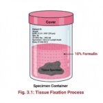
- 10% Formol Saline
- 10% Neutral Bufered Formalin
- Heidenhain’s Susa
- Formol sublimate
- Zenker’s solution
- Zenker formol
- Ouin’s solution
- Brasil’s solution
B. Nuclear Fixative – Preserve nuclear structures such as,
- Flemming’s fluid
- Carnoy’s fluid
- Bouin’s fluid
- Newcomer’s fluid
- Heidenhain’s Susa
C. Cytological Fixatives – preserves cytoplasmic structures such as,
- Flemming’s fluid without acetic acid
- Kelly’s fluid Formalin with “post-chroming”
- Regaud’s fluid (Muller’s fluid)
- Orth’s fluid
- Histochemical Fixatives – preserve chemical contents of cells and tissues such as
D. LIPID FIXATIVE – Mercuric chloride and Potassium dichromate
- PHOSPHOLIPIDS FIXATIVE – Baker’s formal calcium
- CARBOHYDRATE FIXATIVE – Alcoholic formaldehyde
- PROTEIN FIXATIVE – Neutral buffered formal saline or formaldehyde
- GLYCOGEN FIXATIVE – Rossman’s fluid or absolute alcohol
Composition, Advantage, Disadvantage & Use of Fixative
- Formaldehyde –
A. 10% formaline
widely used (10% formalin)
Disadvantage –
- fumes are irritating to the nose and eyes
- prolonged storage may induce precipitation of white paraformaldehyde
Notes – Removal of precipitate is addition of 10% methano
B. 10% formol – Saline –
- – 40% Formaldehyde + NaCl + Distilled water
- fixation of CNS Tissues and General post-mortem tissues
- preserves enzymes and proteins
C. 10% Neutral Buffered Formalin/Phosphate-Buffered Formalin –
- Sodium dihydrogen phosphate + Disodium hydrogen phosphate + 40%Formaldehyde + Distilled water
- Preservation of surgical, post-mortem and research specimens
- Best fixative for iron-containing tissues
D. Formol-Corrosive (Formol Sublimate)
- Aq. Mercuric Chloride + 40% Formaldehyde
- Routine post-mortem tissues
- Excellent in silver reticulum methods
- Fixes lipids, especially neutral fats and phospholipids
E. Alcoholic Formalin (Gendre’s Fixative)
- 95% Ethyl Alcohol saturated with picric acid + Strong formaldehyde solution + glacial acetic acid.
- Immunoperoxidase studies on tissues
- Used for rapid diagnosis
- Good for preservation of glycogen and for micro-incineration
- Used to fix sputum, since it coagulate mucus
F. Glutaraldehyde
- two formaldehyde residues linked by 3C chains
- used for enzyme histochemistry and electron microscopy
- preserves plasma proteins
2. METALLIC FIXATIVES
A. MERCURIC CHLORIDE
- Mercuric Chloride + Potassium Dichromate + Sodium Sulfate + Distilled Water
- most common metallic fixative
- Tissues fixed with mixtures containing mercuric chloride (except Susa) contain black precipitates of mercury.
- Routine fixative of choice for preservation of cell detail in tissue photography.
- Renal tissues, Fibrin, Connective tissues and muscles
- Black deposits may be removed by adding saturated iodine solution in 96% alcohol, the iodine being decolorized with absolute alcohol in the subsequent stages of dehydration.
B. Zenker’s Fluid
- Mercuric Chloride + Glacial Acetic Acid
- fixing small pieces of liver, spleen, connective tissue and nuclei
- may act as mordant
- Mercuric deposits may be removed by immersing tissues in an alcoholic iodine solution. “de-zenkerization”
C. Zenker-formol (Helly’s solution)
- Mercuric chloride + Potassium dichromate + Sodium sulphate + Distilled water + Strong formaldehyde (40%)
- Fixative for pituitary gland, bone marrow and blood-containing organs such as spleen and liver.
- Preserves cytoplasmic granules
- Brown pigments are produced if tissues are allowed to stay for more than 24 hours.
- Pigments can be removed by immersing the tissue in saturated alcoholic picric acid or sodium hydroxide
D. Heidenhain’s Susa Solution
- Mercuric chloride + Sodium chloride + Trichloroacetic acid + Glacial Acetic Acid + Formaldehyde (40%) + Distilled water
- tumor biopsies especially of the skin
- Excellent cytological fixative
- Mercuric chloride deposits may be removed by immersion on alcoholic iodine solution
- the tissue should be transferred directly to a high-grade alcohol, to avoid undue swelling of tissues caused by treatment with low-grade alcohol or water.
E. B-5 Fixative
- Distilled water + Mercuric Chloride + Sodium acetate
- Commonly used for bone marrow biopsies