


STAINING OF SPIROCHETES
Introduction-
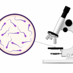
The three important groups of spirochetes are (1) Treponema (2) Leptospira and (3) Borrelia. These are motile, elongated, flexible and spiral organisms. They are not easily stained and can be demonstrated by silver impregnation method (Fontana’s method) or by dark field microscopy.
1. Treponema: These are about 0.2 μm in width and 5 to 15 μm in length.
1. Treponema pallidum: This is the causative or- ganism of syphilis which is transmitted by sex- ual contact.
2. Treponema pertenue: In morphology it is iden- tical with Treponema pallidum and it is the caus- ative organism of yaws. It causes ulcerating papule on the arms or legs.
3.Treponema carateum: It causes nonulcerative papule and leads to the disease Pinta (predom- inantly occurs in Mexico).
4.Treponema microdentum: This organism is found in the secretions between the teeth and in the tartar.
5. Treponema calligyrum: These organisms may occur in genital secretions.
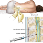
2. Leptospira: These vary from 5 to 15 um in length and about 0.1 um in width. Leptospira icterohem- orrhagiae causes Weil’s disease (infectious jaundice). It is transmitted through water or food contaminated through animal sources (infected rats, mice or dogs). Leptospira can be detected in blood by the dark field illumination.
3. Borrelia: These are irregular wide open coils and are larger in length (10 to 30 μm). These can be stained with aniline and Romanowsky stains.
STAINING OF SPIROCHETES
Principle-
Since heat denatures the spirochetes, a chemical fixative such as absolute alcohol is used as a fixative. Afterwards silver impregnation method of Fontana is used. Deposition of silver salt in the organism makes them visible microscopically.
Reagents-
1. Fixative
A) Glacial acetic acid 1 ml
b) Formalin 2 ml
c) Distilled water to 100 ml
2. Mordant
a Phenol 1g
b) Tannic acid 5g
c)Distilled water to 100 ml
3 .Ammoniated silver nitrate
a) 10% (v/v) ammonia. b) 0.5% (w/v) silver nitrate.
4. Normal saline: (0.85% (w/v) sodium chloride in distilled water.
Note-
In the case of solution 3, add solution (a)
To solution (b) Until the precipitate formed just dissolves. Add solution (b)
Dropwise till the precipitate returns. The precipitate should not redissolve
again.
Procedure-
1. Make a smear of tooth tartar in
a drop of normal saline.
2. Treat the film three times (30
seconds each) with the fixative.
3. Wash off with absolute alcohol
and flood with alcohol for about 3 minutes.
4. Drain alcohol completely and
air dry the smear.
5. Flood the smear with mordant.
Heat it till steam rises for 30 seconds.
6. Wash the smear under running
tap water. Dry the smear completely.
7. Treat with ammoniated silver
nitrate. Heat till steam rises for 30 seconds (The film should appear brown).
8. Wash in distilled water and dry
in air,
9. Mount in Canada balsam under
coverslip (since some immersion oils cause the film to fade).
10. Observe under oil immersion
lens.
Result-
a) Spirochetes Browinsh black
b) Background Brownish yellow