


GRAM STAIN :-
Objective :-
To differentiate bacteria into Gram-positive and Gram-negative based on the ability of their cell wall to retain the primary stain (crystal violet) after decolorization.
Principle
- Gram-positive bacteria: Thick peptidoglycan layer →
retain crystal violet–iodine complex → appear purple.
- Gram-negative bacteria: Thin peptidoglycan, high lipid content → lose crystal violet on decolorization → take up safranin → appear pink/red.
Materials Required
- Clean glass slides
- Inoculating loop / needle
- Bunsen burner
- Staining rack
- Wash bottle with water
- Blotting paper
Reagents
- Crystal Violet (Primary stain)
- Gram’s Iodine (Mordant)
- Decolorizer (Acetone–alcohol or 95% ethanol)
- Safranin (Counterstain)
Procedure
1. Preparation of Smear
- Clean the slide and label it.
- Place a small drop of water on the slide.
- Pick a small amount of culture and spread to form a thin smear.
- Air dry completely.
- Heat fix by passing the slide over flame 2–3 times (do not overheat).
· 2. Gram Staining Steps
Step | Reagent | Time |
|
1 | Crystal Violet | 1 minute |
|
2 | Wash gently | — |
|
3 | Gram’s Iodine | 1 minute |
|
4 | Wash gently | — |
|
5 | Decolorizer (Alcohol/Acetone) | Few seconds (5–15 sec) |
|
6 | Wash immediately | — |
|
7 | Safranin | 30–60 seconds |
|
8 | Final wash | — |
|
9 | Blot dry | — |
|
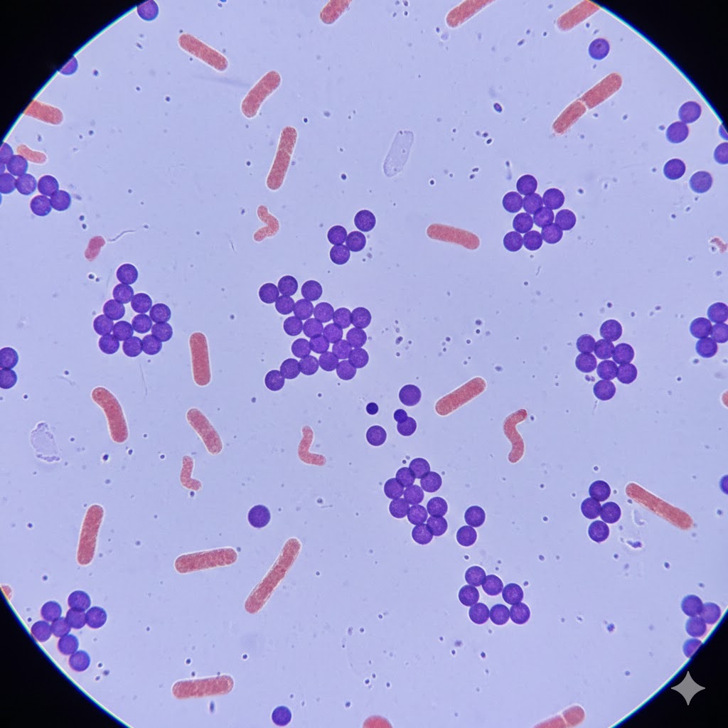
Microscopic Examination
- Observe under oil immersion (100x) objective.
Findings
- Gram-positive → Purple / violet
- Gram-negative → Pink / red
- Note shape: cocci, bacilli, spirilla, clusters, chains, pairs.