


Introduction:-
Special Light Microscope refers to advanced microscopic techniques used to examine specimens that cannot be clearly observed under a simple compound microscope. Microscopes are instruments designed to produce magnified visual or photographic images of objects too small to be seen with the naked eye. A microscope must accomplish three tasks: produce a magnified image of the specimen, separate the details in the image, and render those details visible to the human eye or camera. Compound microscopes are suitable for examination of stained preparations, but for special conditions we require a Special Light Microscope such as dark-ground, phase contrast, polarizing, and immunofluorescence microscopes.
Objectives of Special Light Microscope
After reading this lesson, you will be able to: *describe the principle of Dark-ground, phase contrast, polarizing and
immunofluorescence microscopy. *explain the uses of Dark-ground, phase contrast, polarizing and
immunofluorescence microscopy.
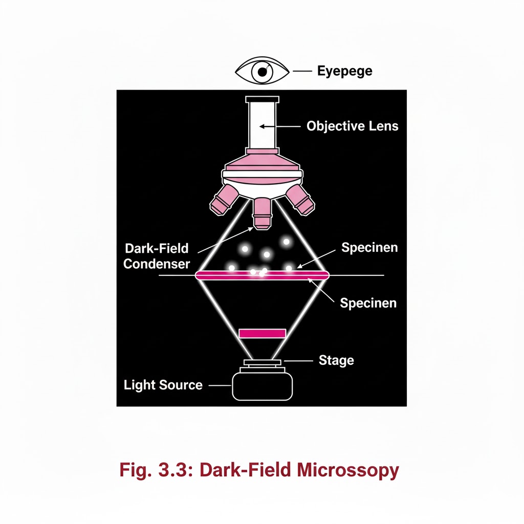
DARK GROUND ILLUMINATION:- Conditions arise when specimen need to be visualized as unstained or living cells. Since such specimens have refractive indices close to medium in which they are suspended, bright field microscopy is difficult as there is not much contrast.
Principle:- Dark ground microscopy prevents direct light from entering the front
of the objective, only light which enters is which gets reflected or diffracted by the specimen, thus making them appear bright in a dark background .
Uses of Dark ground microscopy::-
Useful for spirochaetes, flagellates, cell
suspensions, flow cell techniques, parasites, autoradiography, and fluorescence microscopy.
Disadvantage: Resolution is inferior to bright field microscopy. Does not reveal
internal details
PHASE CONTRAST MICROSCOPY:-
Unstained and living biological material viewing by bright field and dark ground
illumination has problems of reduced illumination and resolution. To overcome
these problems, phase contrast microscopes are used.
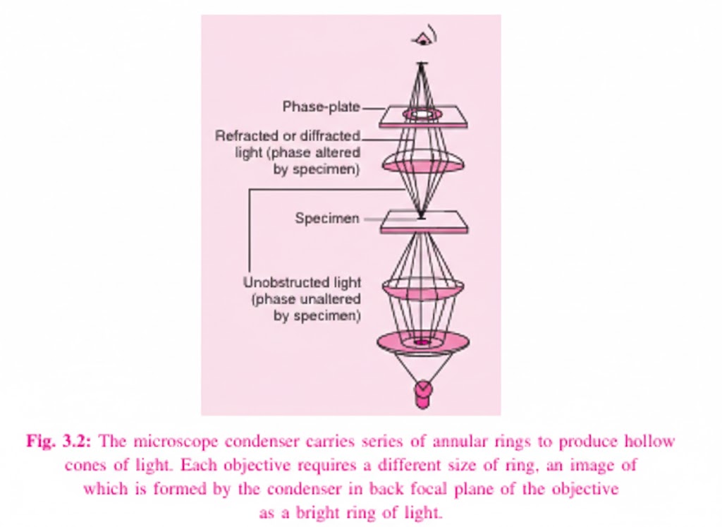
Principle: It is an optical microscopy illumination technique that converts phase shifts in light passing through a transparent specimen to brightness changes in the image. The phase shifts themselves are invisible to the human eye, but become visible when they are shown as brightness changes.
A practical implementation of phase-contrast illumination consists of a phase
ring (located in an aperture plane located somewhere behind the front lens
element of the objective) and a matching annular ring, which is located in the
conjugate primary aperture plane
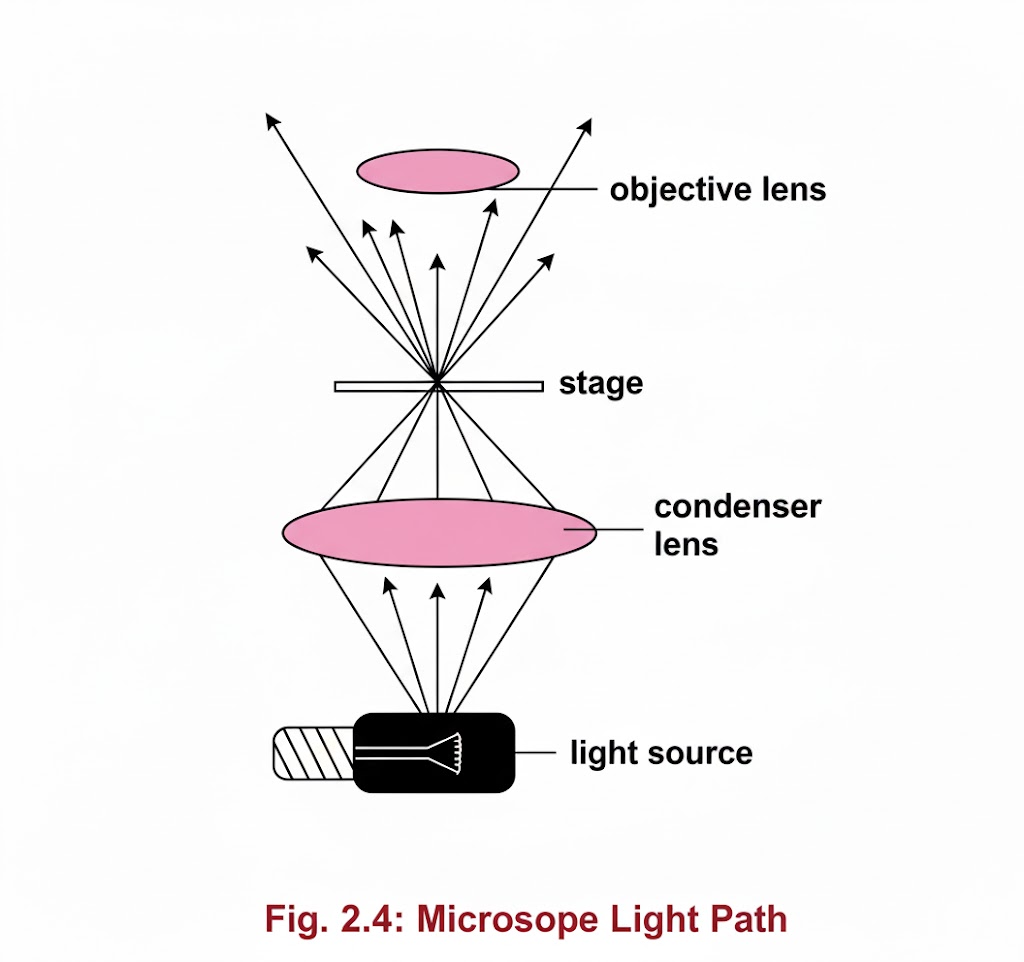
Two selected light rays, which are emitted from one point inside the lamp’s
filament, are focused by the field lens exactly inside the opening of the condenser
annular ring. Since this location is precisely in the front focal plane of the condenser, the two light rays are then refracted in such way that they exit the condenser as parallel rays. Assuming that the two rays in question are neither refracted nor diffracted in the specimen plane (location of microscope slide),
they enter the objective as parallel rays. Since all parallel rays are focused in the back focal plane of the objective, the back focal plane is a conjugate aperture
plane to the condenser’s front focal plane (also location of the condenser annulus). To complete the phase setup, a phase plate is positioned inside the back focal plane in annulus
Uses: It’s a quick and efficient way of examining unstained paraffin, resin and
frozen sections, studying living cells (cell cultures) and their behavior.
POLARIZED LIGHT MICROSCOPY:-
Light can be described as an electromagnetic vibration where there are many planes of vibration. Natural light vibrates in many planes or directions, whereas polarized light vibrates in only one plane. It can be produced by passing light through a polarizer. Substances capable of producing polarized light are called birefringent.
Principle: The dedicated polarizing microscope uses two polarizers. One,
always referred to as polarizer, is placed beneath the substage condenser. The
other is called analyzer and is placed between the objective and the eyepiece.
Looking through both polarizers, the light intensity is best when they are both giving light vibrating parallel to each other. However, when the light vibration is at right angles to each other, there is dark background. If a substance capable
of birefringence is placed between the two polarisers it gets visualized as brightness against a black background.
Uses: Medicine-Amyloid detection, collagen fibers, urates and other crystals.Metallurgy, Ceramics.
FLUORESCENCE MICROSCOPY:-
Principle: Fluorescence is the property of some substances which, when illuminated by light of a certain wavelength, will re-emit the light at a longer wavelength. In fluorescence microscopy, the exciting radiation is usually ultra violet wavelength or blue region.
A substance which possesses a fluorophore will fluoresce naturally (Primary or
autofluorescence) eg Vitamin A, chlorophyll
Dyes, chemicals and antibodies added to tissues produce secondary fluorescence
of structures and are called fluorochromes. When antibodies labeled with
fluorochromes are used to detect particular antigens, the technique is called
Immunofluorescent technique and is widely used in medicine. Tissue antigens
most commonly demonstrable by Immunofluorescence are viruses, protozoa,
bacteria, enzymes, hormones, plasma proteins, cells and cell constituents.
Examples of fluorochromes: Fluorescein (apple green emission color), Rhodamine (Orange-red color