


INTRODUCTION:-
The presence of calcium salts in tissues makes them hard. This causes damage
to the knife, difficulty in cutting tissue. Calcium is normally present in bones
and teeth. Calcium may also be present in normal tissues in pathological
conditions like necrotic tissue in tuberculosis.
OBJECTIVES:-
After reading this lesson, you will be able to:
*describe decalcification
*explain different methods of decalcification
*describe the chemical and physical tests to estimate the remaining calcium.
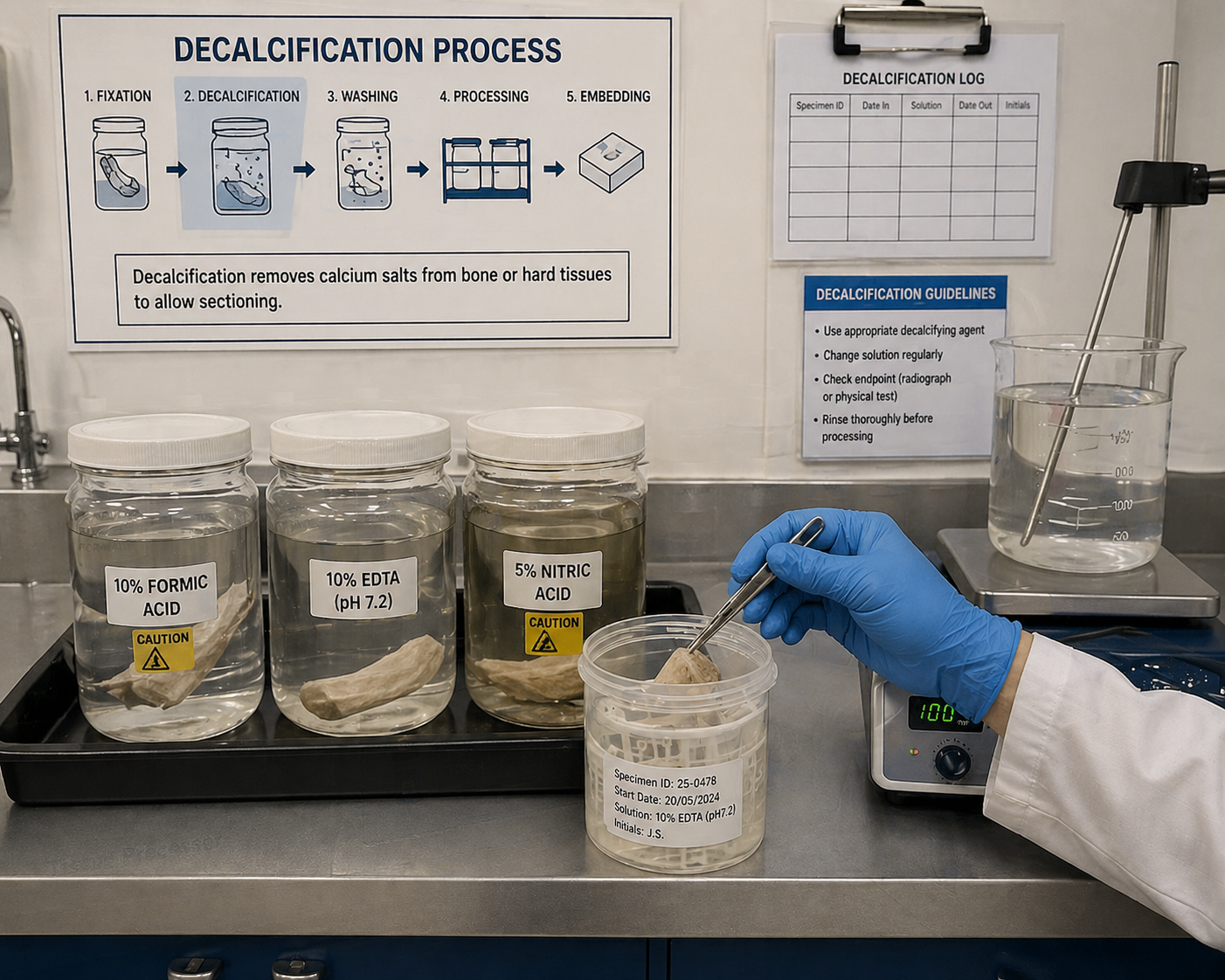
DECALCIFICATION:-
Aim – To remove calcium salts from the tissues and make them amenable for sectioning.
Preparation of tissues – The calcified hard tissues should be first cut into small pieces (2 to 6mm) with a thin blade, hacksaw or sharp knife in order to minimize the tearing of the surrounding tissues. This process is followed by fixation in buffered formalin or any other desired fixative. After fixation tissues must be thoroughly washed and excess fixative should be removed before the specimen is subjected to decalcification.
DIFFERENT METHODS OF DECALCIFICATION:-
1. Acid decalcification
2. Ion exchange resin
3. Electrical ionization
4. Chelating methods
5. Surface decalcification
Decalcification process should satisfy the following conditions
*Complete removal of calcium salts
*Minimal distortion of cell morphology
*No interference during staining
Decalcification is a straightforward process but to be successful it requires:
*A careful preliminary assessment of the specimen
*Thorough fixation
*Preparation of slices of reasonable thickness for fixation and processing
*The choice of a suitable decalcifier with adequate volume, changed
regularly
*A careful determination of the endpoint
*Thorough processing using a suitable schedule.
Methods of Decalcification :-
The tissue is cut into small pieces of 3 to 5 mm size. This helps in faster decalcification. The tissue is then suspended in decalcifying medium with waxed thread. The covering of wax on thread prevents from the action of acid on thread. The volume of the decalcifying solution should be 50 to 100 times of the volume of tissue. The decalcification should be checked at the regular interval.
Acid Decalcification –
This is the most commonly used method. Various acid solutions may be used alone or in combination with a neutralizer. The neutralizer helps in preventing the swelling of the cells.
Following are the usually used decalcifying solutions-
1. Aqueous Nitric Acid:-
Nitric acid – 5ml
Distilled water – 100 ml
If tissue is left for long time in the solution, the tissue may be damaged. Yellow colour of nitric acid should be removed with urea. But this solution gives good nuclear staining and also rapid action.
2. Nitric Acid Formaldehyde
Nitric acid – 10 ml
Formaline – 5-10 ml Distilled water upto 100 ml
Advantages
* Rapid action
*Good nuclear staining
*Washing with water is not required
*Formalin protects the tissues from maceration
3. Formic Acid Solution:
Formic acid – 5 ml
Distilled water – 90 ml
Formalin – 5 ml
In this solution the decalcification is slow. If concentration of formic acid is increased the process is fast but tissue damage is more:
4. Trichloroacitic Acid – This is used for small biopsies. The process ofdecalcification is slow hence cannot be used for dense bone or big bony pieces.
Formal saline (10%) – 95 ml
Tricloroacitic acid – 5 gm
Ion Exchange method –
In these ammonium salts of sulfonated polystyrene resin is used. The salt is layered on the bottom of the container and formic acid containing fluid is filled. The decalcifying fluid should not contain mineral acid. X-rays can only determine complete decalcification. The advantages of this method are:-
*Faster decalcification
*Well preserved tissue structures
*Longer use of resin
Electrolytic Method – Formic acid or HCl are used as electrolytic medium. The
calcium ions move towards the cathode. Rapid decalcification is achieved but
heat produced may damage the cytological details.
Chelating Agents:- Organic chelating agents absorb metallic ions. EDTA can
bind calcium forming a non-ionized soluble complex. It works best for
cancerous bone. This is best method for decalcification of bone marrow biopsies
as it preserves cytological details best. The glycogen of marrow is preserved.
EDTA Solution :-
EDTA – 5.5 gm
Formaline – 100 ml
Distilled water- – 900 ml
Surface Decalcification – The surface layer of paraffin blocks are inverted in5% HCl for one hour. About top 30 micron is decalcified. It should be washed thoroughly before cutting.
Factors affecting rate of Decalcification:-
1. Concentration of decalcifying solution-Increased concentration of the
decalcifying agent fastens the reaction.
2. Temperature-The rate of decalcification increases with rise of temperature.
3. Density of bone-Harder bone takes longer time to decalcify.
4. Thickness of the tissue-Small tissue pieces decalcify earlier.
5. Agitation-Agitation increases the rate of decalcification.
METHODS OF DETERMINING OPTIMUM DECALCIFICATION OR ENDPOINT:-
Specimens should NOT be crowded together and should NOT contact the bottom of container in order to provide complete decalcification. Over decalcification can also permanently damage specimen. The following procedure help determine the correct end-point of decalcification.
End-Point of Decalcification:
X-ray (the most accurate way)
Chemical testing (accurate)
Physical testing (less accurate and potentially damage of specimen)
Chemical Test: The following solutions are needed to chemically test for residual calcium.
5% Ammonium Hydroxide Stock:
Ammonium hydroxide, 28% 5 ml
Distilled water 95 ml
Mix well
5% Ammonium Oxalate Stock:
Ammonium oxalate 5 ml
Distilled water 95 ml
Mix well
Ammonium Hydroxide/Ammonium Oxalate Working Solution:
Use equal parts of the 5% ammonium hydroxide solution and the 5% ammonium
oxalate solution.
Procedure:-
1. Insert a pipette into the decalcifying solution containing the specimen.
2. Withdraw approximately 5 ml of the hydrochloric acid/formic acid
decalcification solution from under the specimen and place it in a test tube.
3. Add approximately 10 ml of the ammonium hydroxide/ammonium oxalate
working solution, mix well and let stand overnight.
4. Decalcification is complete when no precipitate is observed on two
consecutive days of testing. Repeat this test every two or three days.
Physical Tests:-
The physical tests include bending the specimen or inserting a pin, razor, or scalpel directly into the tissue. The disadvantage of inserting a pin, razor, or scalpel is the introduction of tears and pinhole artifacts. Slightly bending the specimen is safer and less disruptive but will not conclusively determine if all calcium salts have been removed. After checking for rigidity, wash thoroughly prior to processing
Note: If paraffin embedded bones are not decalcified fully, one can soak the paraffin blocks in the same decalcification solution for a few minutes before cutting. This is usually helpful.
Points to remember:-
*After completion of decalcification, the specimen should be washed in
water
*Over decalcification is more noticeable in staining of nuclei.
*Acid solutions soften bone by removing calcium salts.
*EDTA is used as chelating agent for decalcification.
*To offset the hydrolysis of nucleic acids caused by decalcification, bone marrow is often fixed in Zenker’s solution.
*During decalcification, carbon dioxide gas is released.
*Factors affecting decalcification are
o Size of specimen,
o Concentration of decalcifying solution, o Time in decalcifying solution o Amount of decalcifying solution
For proper decalcification, bone should be cut into 4-5mm thick pieces
Chelating agents act by binding calcium ions
s