


INTRODUCTION:-
The sections, as they are prepared, are colourless and different components cannot be appreciated. Staining them by different coloured dyes, having affinities of specific components of tissues, makes identification and study of their morphology possible. Hematoxylin and Eosin (H&E) is the most frequently used stain in histology.
OBJECTIVES:-
After reading this lesson, you will be able to: describe Hematoxylin and its preparation describe the properties of Hematoxylin explain Eosin and its preparation describe the method of staining.
HEMATOXYLIN :-
It is extracted from the bark of a tree”, hematoxylom campechianum”. The hematoxylin which we buy is extracted from this bloodwood tree. To obtain the bark of freshly logged tree is chipped off, then boil the chips in water. An orange red solution is obtained, which turns yellow, then black on cooling. The water is evaporated leaving crude hematoxylin. Further purification is done.Solutions of the dye should be oxidized to retain its staining ability longer. The dye may be oxidized by exposure to the natural light for 3-4 months.
chemical oxidation is achieved by using either sodium iodate or mercuric oxide. The chemical oxidation converts the dye almost instantaneously but the product does not have shelf life. Sodium iodate is most commonly used oxidizing agent (0.2gm oxidizes 1.0 gm hematoxylin).
Hematoxylin is neither a dye nor it has coloring properties. For nuclear staining it is necessary to oxidize the hematoxylin to hematin which is a weak anionic purple dye. Anionic hematin will have no affinity for the nucleic acids of nuclei. Hence a metallic salt or mordant is combined with hematoxylin so that a positive charge to the dye is obtained by virtue of the metal action. Thus the cationic dye–metal complex will bind to the anionic nuclear chromatin. Various mordants are ammonium or potassium alum ferric salt, chrom alum and phosphotungstic acid. The tissue component most frequently demonstrated is nuclear chromatin using an alum mordant in the H&E staining method.
The combination of hematoxylin and mordant is called a hematoxylin lake. The aluminium lake formed with ammonium alum is particularly useful for staining nuclei. Hematoxylin recipes using these mordants are called alum hematoxylin.
PROPERTIES OF HEMATOXYLIN :-
1. Hematoxylin has no staining property.
2. Hematin with mordant such as ammonium or potassium alum forms lake
which functions as cationic dye and stains anionic tissue components.
3. Hematin in an aqueous solution can be acidic or an alkaline dye depending
on pH.
4. Hematin has affinity for several tissues with an appropriate mordant.
Progessive staining :-When tissue is left in the stain just long enough to reach the proper end point. The slides have to be examined at different interval to find out when the staining is optimum.
Regressive staining :- In this method the tissue is overstained and then destained (differentiate) until the proper endpoint is reached.
Harris hematoxylin is a regressive stain; the overstaining is removed by acid alcohol. The removal of this excess dye is called differentiation.
The hematoxylin alum gives a reddish hue to the tissues because of acidic pH. To convert this colour to the final blue, alkaline pH is required. This process is called “blueing”. It is done either by tap water or by ammonium hydroxide.
Preparation of Harris’s hematoxylin :-
Ingredients :-
Hematoxylin – 5 gm
Absolute alcohol – 50 ml
Ammonium alum – 100 gm
Distilled water – 1000 ml
Mercuric oxide – 2.5 gm
Glacial acetic acid 5gm – 40 ml
Method :- Dissolve the hematoxylin in absolute alcohol and ammonium alum in hot water. Mix the two solutions and heat to boiling. Remove from flame, and add mercuric oxide and cool rapidly. Glacial acetic acid if added gives brisk nuclear staining, but life of the solution is reduced. Hence if acetic acid is to be added, it should be added in working solution.
Preparation of Mayer’s hematoxylin:-
Ingredients :
Hematoxylin – 1.0 gm Distilled water – 1000 ml Ammonium alum – 50 gm
Sodium iodate – 0.2 gm
Citric acid (reduces pH) – 1.0 gm
Chloral hydrate (preservative)- 50 gm
Method:- Hematoxylin is dissolved in distilled water using gentle heat. Then
alum is added and dissolved. Then sodium iodate, citric acid and chloral hydrate
are added respectively.
EOSIN :-
Eosin is used as the counterstain that stains the cytoplasm rose coloured. The intensity of the eosin is individual choice. The most widely used eosin is “eosin Y”. The “Y” stands for yellowish. It is available in either water soluble or alcohol soluble form. Most laboratories use the water soluble form of eosin Y in an alcohol-water solution which is described here.
Eosin Y (water soluble) – 1.0 gm
Distilled water – 80 ml
95% alcohol – 320 ml
Glacial acetic acid – 0.4 ml
Preparation :- Dissolve eosin in water and then add this to 95% alcohol (one part eosin solution with 4 parts alcohol). To the final mixture add a few drops of acetic acid (0.4ml). The acetic acid increases the staining intensity of eosin. When ready to use, the stain should be cloudy; if clear, add a few drops of the acetic acid. The solution should be standardized by staining the control slides.
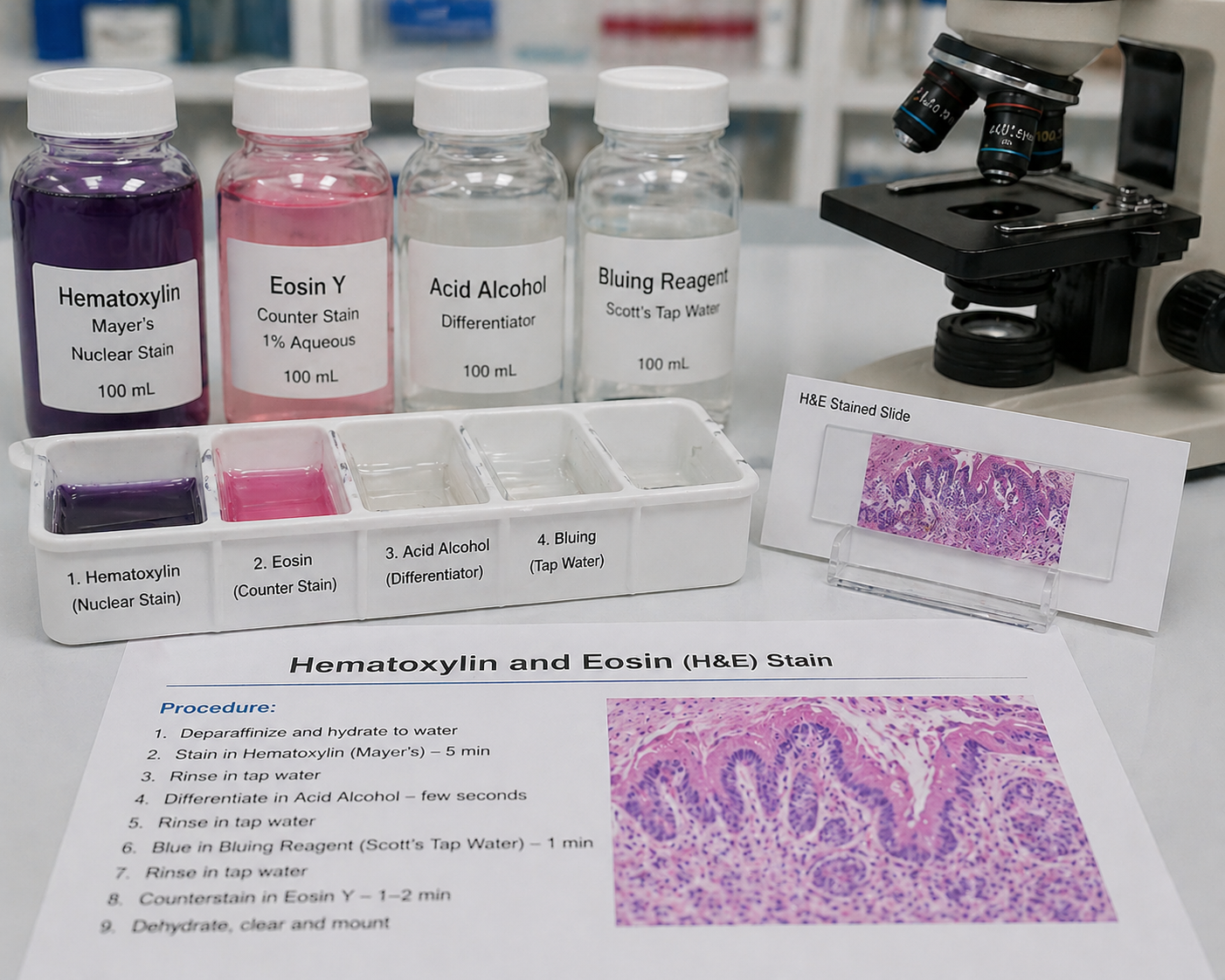
METHOD OF STAINING :-
1. Deparaffinize sections in xylene, 10-20 minutes. Filter Hematoxylin.
2. Rehydrate sections:
100% alcohol for 1-2 minutes
95% alcohol for 1-2 minutes
3. Rinse in tap water
4. Rinse in distilled water
5. Stain with Hematoxylin for 3-5 minutes
6. Wash in tap water
7. Differentiate section with 1% HCl in 70% alcohol 1-2 dips and check under
microscope. If necessary, return slides to HCl for further differentiation.
8. Wash slides in running tape water for 15 minutes
9. Stain slides in Eosin for 1-4 minutes
10. Dehydration and Differentiation:
95% alcohol 5-6 dips
100% alcohol 5-6 dips
11. Clear slides in xylene 2 times
12. Mount slides with mounting media (Permount or DPX)
Note :-
1. At no stage of staining the section should be dry
2. H&E is a regressive stain in which a tissue is over-stained and then excess
dye is removed to obtain desired intensity of stain
3. Filter Hematoxylin each time before staining
4. Change most of alcohol and xylene each time before staining