


INTRODUCTION:-
Sections are prepared quickly for histological examination by freezing the tissue.
The section should be thin, and without water crystals. It is an important
procedure for quick diagnosis.
OBJECTIVES:-
After reading this lesson, you will be able to:
*enlist the indications of frozen section
*explain the disadvantages of frozen section
*describe cryostat
PURPOSES OF FROZEN SECTION :-
Frozen sections are used for following purpose:
*Quick diagnosis
*Study the margins of cancer
*Enzyme histochemistry
*Immunohistochemistry
*Detection of lipid
*Some molecular procedures
Disadvantages:-
*Morphology is distorted
*Cellular details are not well seen,
*Staining is not very good
*Some specials stains cannot be performed.
Handling of specimen:-
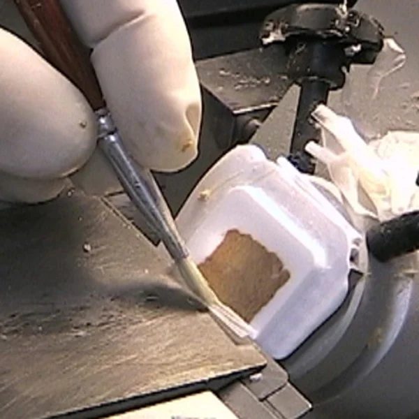
Tissue must reach histopathology laboratory immediately. To avoid tissue being dried it should be kept in saline. The size of the tissue should be small thin, so that good smooth sections can be obtained and freezing is quick. Thickness of he tissue should be about 3mm to 4mm.The tissue can directly be taken to
cryostat or can be fixed with 10% formalin or formol –alcohol
Embedding media:-
Sucrose (20%) or a drop of water may be applied on the chuck. Optimum Cooling temperature (OCT) compounds or 20% sucrose gives good result. Other embedding media are available with cryostat. Completion of freezing is observed by the change of color of tissue which turns glossy white. Freezing should be done fast. This will prevent ice crystal formation. The morphology is better preserved and artifacts are lesDifferent freezing substances are used depending upon the availability and
feasibility.
Carbon Dioxide gas is most commonly used with freezing microtome. This gives good results. Liquid Nitrogen is another substance used for freezing the tissue. An expertise is required while using liquid nitrogen to get uniform freezing. Aerosol sprays are also used for this purpose
Cryostat: Cryostat is used in medicine to cut histological sections. They are usually used in a process called frozen section histology. The cryostat is essentially an ultrafine “deli-slicer”, called a microtome, placed in a freezer. The cryostat is usually a stationary upright freezer, with an external wheel for rotating
the microtome. The temperature can be varied, depending on the tissue being cut – usually from minus 20 to minus 30 degree Celsius. The freezer is either powered by electricity, or by a refrigerant like liquid nitrogen. Small portable cryostats are available and can run off generators or vehicle inverters. To minimize unnecessary warming all necessary mechanical movements of the microtome can be achieved by hand via a wheel mounted outside the chamber. Newer microtomes have electric push button advancement of the tissue.
The precision of the cutting is in micrometres. Tissue are sectioned as thin as 1 micrometre. Usual histology slides are mounted with a thickness of about 7 micrometres. Specimens that are soft at room temperature are mounted on a cutting medium (often made of egg white) on a metal “chuck”, and frozen to cutting temperature (for example at -20 degrees C). Once frozen, the specimen on the chuck is
mounted on the microtome. The crank is rotated and the specimen advances toward the cutting blade. Once the specimen is cut to a satisfactory quality, it is mounted on a warm (room temperature) clear glass slide, where it will instantaneously melt and adhere. The glass slide and specimen are air dried, and
stained. The entire process from mounting to reading the slide takes from 10 to 20 minutes, allowing rapid diagnosis in the operating room, for the surgical excision of cancer. The cryostat section quality is poorer as compared to fixed tissue sections