


SKELETON SYSTEM :-
INTRODUCTION OF SKELETON SYSTEM:–
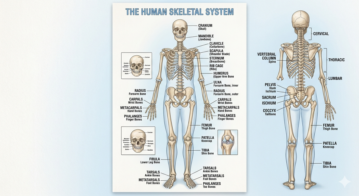
The skeletal system forms the framework of the body. It is the body system composed of bones, cartilage, and ligaments. Each bone serves a particular function and varies in size, shape, and strength.The skeleton consists of the bones of the body. For adults, there are 206 bones in the skeleton. Its providing support and movements of the body It protects the internal organs, including the brain, spinal cord, heart, lungs, and pelvic organs.
The skeleton is subdivided into two major divisions: –
1.Axial Skeleton (80 bones):- Longitudinal axis of the body, including the skull, vertebral column, and thoracic cage (ribs/sternum).
2.Appendicular Skeleton (126 bones): Bones of the limbs and the girdles (pectoral and pelvic)
BONES:-
Bones are a vital component of the vertebrate skeletal system, providing structure and support to the body while also serving crucial roles in mobility, protection, and mineral storage. bones are essential for protecting critical internal organs. For example, the skull shields the brain,the ribcage safeguards the heart and lungs, and the vertebrae encase the spinal cord.
Functions of the bone:-
The function of bones included:-
*providing the body framework
*Giving attachments to muscles and tendons.
*Allowing movement of the body as a whole and of parts of the body , by forming joints that are moved by muscles.
*Haemopoiesis ,the production of blood cells in red bone marrow.
* Mineral storage,especially calcium phosphate- the mineral reservoir within bone is essential for maintenance of blood calcium level, which must be tightly controlled.
*Forming the boundaries of the cranial, thoracic and pelvic cavities,and protecting the organs.
Types of Bones:-
| TYPES OF BONE | SHAPES | EXAMPLE | FUNCTIION |
|---|
| LONG | LONG & CYLENDRICAL | FEMUR | MOVEMENT |
| SHORT | CUBE- LIKE | CARPALS | STABILITY |
| FLAT | THIN & FLAT | SKULL | PROTECTION |
| IRREGULAR | COMPLEX | VERTEBRAE | SUPPORT |
| SESAMOID | SMALL & ROUND | PATELLA | TENDON PROTECTION |
1. AXIAL SKELETON
2. APPENDICULAR SKELETON
1. Axial skeleton:-
The axial skeleton is the central core of the human body ,comprising 80 bones that protect vital organs and support upright posture. its consist of the skull,vertebral column ,sternum and ribs.
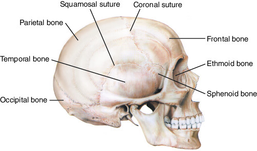
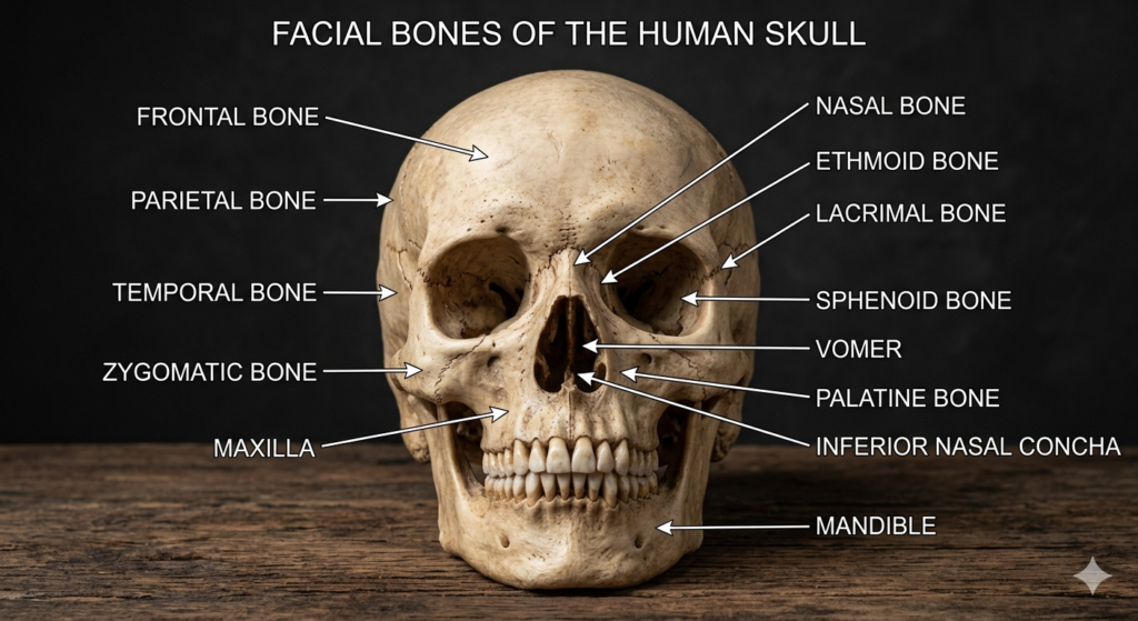
* Skull (22 bones):- 8 cranial,14 facial bone.
- Hyoid Bone (1 bone): U-shaped neck bone that supports the tongue and larynx.
- Vertebral Column (26 bones): 7 cervical, 12 thoracic, 5 lumbar vertebrae, 1 sacrum, and 1 coccyx (tailbone).
- Thoracic cage(25 bones):- 24 ribs(12 pairs) and sternum (breastbone)
- Frontal bone (1)
- Parietal bones (2)
- Temporal bones (2)
- Occipital bone (1)
- Sphenoid bone (1)
- Ethmoid bone(1)
2. facial bone:-
- Nasal bones (2)
- Maxillae (2)
- Zygomatic bones (2)
- Palatine bones (2)
- Lacrimal bones (2)
- Inferior nasal conchae (2)
- Vomer (1)
- Mandible (1)
:-function of skull:–
The various parts of the skull have specific and different functions:-
*The primary function is protecting the brain, cranial nerves, and meninges from injury.
* It gives the face its shape and provides a framework for soft tissues, such as the nose and mouth.
*It forms the boundaries of the nasal cavity for breathing and the oral cavity for eating.
*The teeth are embedded in the mandible and maxilla; and movement of the mandible allows chewing.
*The skull features specialized cavities that house and protect the sensory organs (eyes in orbits, ears, nose).
Vertebral column:-
The vertebral column—also known as the spine, backbone, or spinal column is a flexible,segmented bony structure extending from the base of the skull to the tailbone..its the central axis of the human skeleton consists of 33 vertebrae in children, which fuse during development into 26 distinct bones in adults. Its primary roles are to protect the spinal cord, support the head and upper body weight, and provide the flexibility needed for various movements.The bodies of the bones are separated from each other by intervertebral discs, its described in five parts:-
- Cervical vertebrae (7)
- Thoracic vertebrae (12)
- Lumbar vertebrae (5)
- Sacrum(5 fused)
- Coccyx(4 fused)
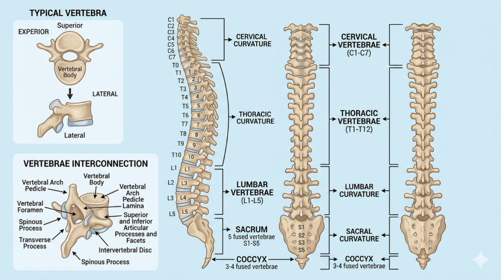
The first vertebra called the atlas, formsa joint (articulates) with the skull.each vertebra forms a joint with the vertebrae immediately above and below.movement are possible in the cervical and lumber regions than in the thoracic region.
The sacrum consists of five vertebrae fused into one bone that articulates with the fifth lumbar vertebra above and coccyx below an innominate (pelvic or hip) bone at each side.
The coccyx consist of the four terminal vertebrae fused into a small triangular bone that articulates with the sacrum above.
Function of vertebral column:-
The vertibral column has several important functions:-
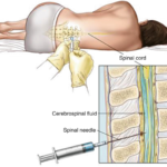
It protect the spinal cord.each vertebra has hole(vertebral foramen) and collectively the foramina form a canal in which the spinal cord lies.
Adjacent vertebrae form openings (intervertebral foramina) which protect the spinal nerves as they pass from the spinal cord
In the thoracic region the ribs articulate with the vertebrae forming joints that allow movement of the ribcage during respiration.
It provides the main support for the head and trunk. Helps maintain an upright posture.
Enables bending, twisting, and rotation of the body. Works with muscles and joints for flexibility.
Act as a shock absorbers Intervertebral discs between vertebrae absorb shocks during walking, running, and jumping.
Transfers body weight from the head and trunk to the pelvis and lower limbs.
Thoracic cage :-
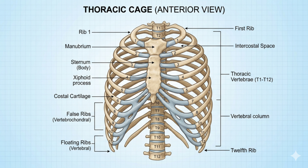
The thoracic cage (also known as the rib cage) is a domed, bony framework that forms the chest portion of the body. its consist of 12 thoracic vertebrae, 12 pairs of ribs with costal cartilages, and the sternum.Its protects vital thoracic organs(heart,lungs)support the upper body,and enables breathing through expansion and contraction acting as a crucial anchor for respiratory and pectoral muscles.
Ribs:-
Ribs cage is the cage-like structure of bones that frames chest cavity (thoracic cavity)it also called thoracic cage.The ribs cage is composed of multiples bones that wrap around the thoracic cavity. the ribs are crucial in the protection of the heart and lungs and help in breathing.Classified by their sternal attachment, they include true, false, and floating ribs, plus structural categories: typical and atypical.
Types of ribs:-
1.True Ribs (Pairs 1–7):- vertebrosternal ribs, are called “true” ribs because. these are connect directly to the sternum through their own individual costal cartilage.
2.False ribs(vertebrochondral ribs):- Pairs 8–10 are called “false ribs”because they don’t connect directly to sternum,they attached to front of ribe cage by the interchondral joints.
3. Floating ribs(vertebral ribs or free ribs) :- Pairs 11–12 are called floating ribs these do not connect to the sternum at all, attaching only to the vertebrae at the back and ending in the abdominal muscles.
Structural Classification of ribs:-
1.Typical Ribs (3rd–9th):- These share a common structure: a wedge-shaped head (with two facets for vertebral articulation), a neck, and a curved body.
2. Atypical ribs(1st,2nd,10th,11th,12th):-These have variations in structure(e.g the 1st is short/flat,while 11th/12th have one facet).
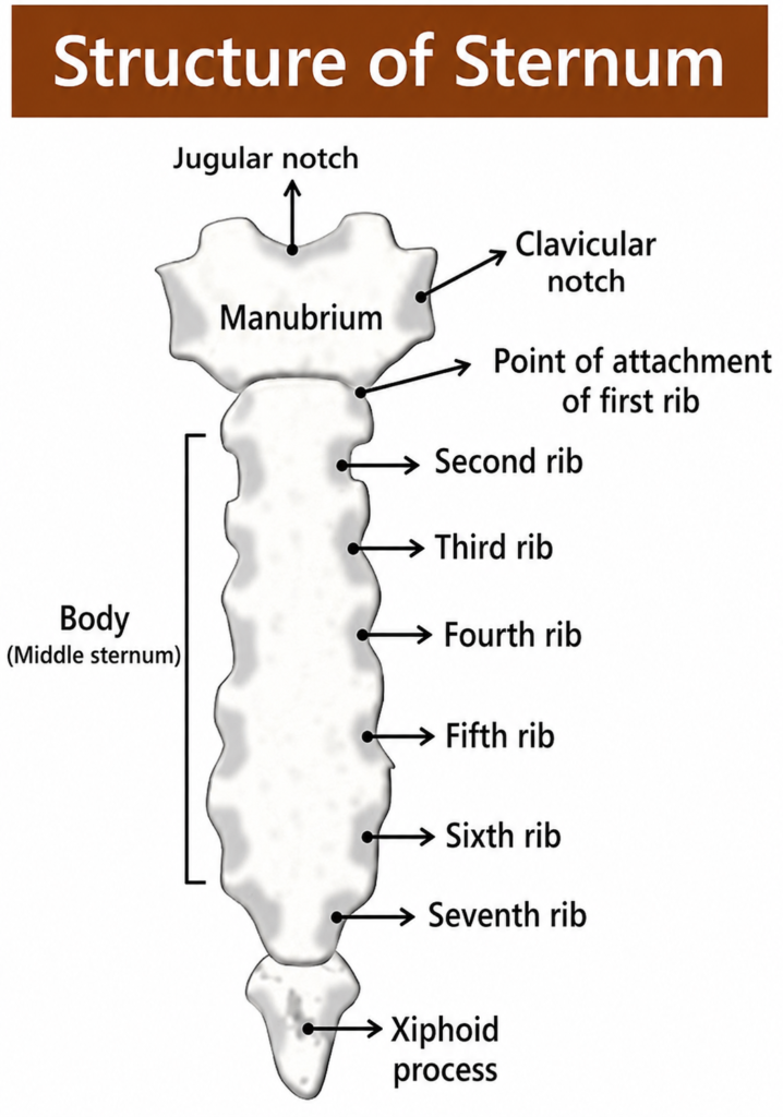
Sternum:-The sternum commonly known as breastbone,is a long,flat,T-shaped bone situated in the center of the chest. it protects the thoracic organs like heart,lungs and major blood vessels serves as the anterior anchor for the ribcage.
its classified structurally as a flat bone and functionally as a part of axial skeleton.its composed of three main part which fuse togather:-
1.Manubrium:-Manubrium is the uppermost and broad part of the sternum has a superior border with a central jugular (suprasternal) notch.on each side of superior border is a clavicular notch for the articulate with the clavicle (collarbones).at the lower side facets on each side for articulate with first costle cartilages.
2.Body (Gladiolus):-body is the middle part of the sternum its long,flat part below the manubrium. its has seven pairs of costle notche on each side for articulates with costle cartilage of ribs 2 to 7.its provide attachment to the cartilage of the ribs form the anterior part of thoracic cage.
3.Xiphoid Process:- Small pointed and variable part at the inferior end its made of hyaline cartilage.its serves as attachment site for the diaphragm and rectus abdomines muscles.
A
Function of thoracic cage:-:-
*The thoracic cage protect the contents of the thorax inculding the heart,lungs and large blood vessels.
*Its forms joint between the upper limbs and the axial skeleton .(upper part of sternum (manubrium) articulates with the clavicles form joint between the upper limbs and the axial skeleton.)
*The thoracic cage gives attachments to the muscles of repiration:- intercostal muscles present between the ribs when they contract the ribs move upwards and outwards,increasing the capacity of the thoracic cage . (The diaphragm is a dome-shaped muscles which separates the thoacic and abdominal cavity )when it contracts it assists with inspiration and experiation .
*Its helps us breathe.
*It acts as the core of the axial skeleton, supporting the shoulder girdle and protecting against the negative pressure created inside the lungs during inhalation.
APPENDICULAR SKELETON :-
The appendicular skeleton is coposed of the bones of the upper limbs ,lower limbs ,shoulder girdle, that attaches the upper limbs and pelvic girdle that attaches to lower limbs of the body.its primary role is facilitate movement and locomotion.
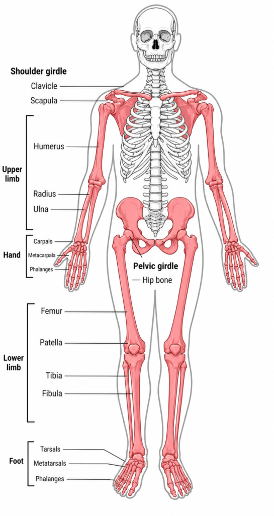
he appendicular skeleton is divided into four main regions:-
1.Pectotal girdles:- The pectoral girdle bone are provided the point of attachment of the upper limbs to the axial skeleton.its consist of the clavicle (collarbone) in the anterior and the scapula ( shoulder blades )in the posterior side.
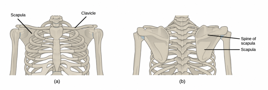
Clavical(2) :-The clavical bone is a long ,curved bone which horizontally articulate wirh scapula and sternum at the frunt of the thorax(chest) just above the first ribs.its S shaped bone position in the arms of body and connected the upper limbs .
Scapula(2) :- Scapula are flat,triangular bones that are located at the posterior thoracic wall of the pectoral girdle,they support the muscles of shoulder joints,its articulate with clavicle and humerus. its play a importent role in muscles attachment &shoulder socket.
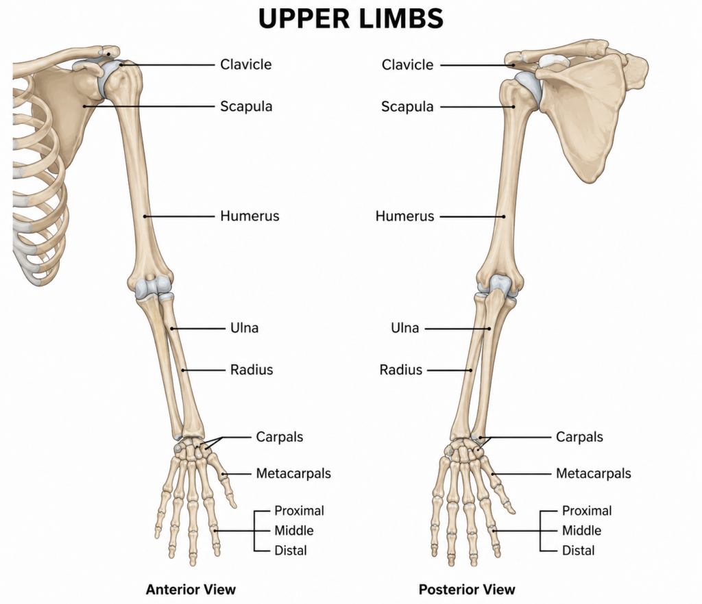
2.upper limbs:-The upper limb contains 30 bones in one side four regions:
1.Shoulder girdle:-
*Clavical(1)(collarbone) *Scapula(1)(shoulder blades)
2.Arms:- Its contain only one bone.
*Humerus(1):-is the largest and longest bone of the upper limb and the only bone of the arm. It articulates with the scapula at the shoulder and with the forearm at the elbow.
3.Forearm:-The forearm extends from the elbow to the wrist it contain two bone Radius & ulna.
Radius(1) is located along the lateral(Thumb) side of forearm and articulates with the humerus at the elbow. Ulna(1) is located on the midial aspect(small finger side) of the forearm,articulates with the humerus.its longer than the radius.
4.Wrist and hand:-its contain 27 bones
*Carpus(wrist) consist eight (8) bone. *Metacarpus(palm)consist 5 bone. *Phalanges(finger)consist 14 bones each finger consist three phalanges except thumb(only two phalanges).
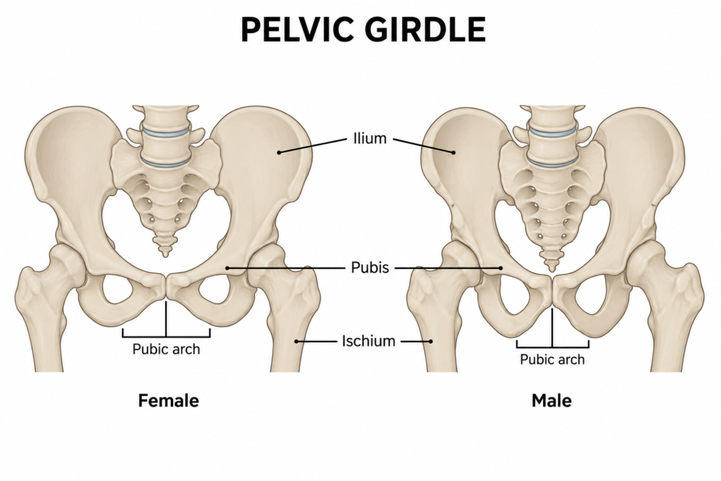
3.Pelvic girdle(2):- The pelvic girdle attaches to the lower limbs of the axial skeleton. Because it is responsible for bearing the weight of the body and for locomotion, the pelvic girdle is securely attached to the axial skeleton by strong ligaments. It also has deep sockets with robust ligaments to securely attach the femur to the body. The pelvic girdle is further strengthened by two large hip bones. In adults, the hip bones, or coxal bones, are formed by the fusion of three pairs of bones: the ilium, ischium, and pubis.
The female pelvis is slightly different from the male pelvis. Over generations of evolution, females with a wider pubic angle and larger diameter pelvic canal reproduced more successfully. Therefore, their offspring also had pelvic anatomy that enabled successful childbirth.
Lower limbs:-The lower limb consists of 30 bone in one side divided in three regions:-Thigh, Leg, and Foot.The bones of the lower limbs are thicker and stronger than the bones of the upper limbs because of the need to support the entire weight of the body and the resulting forces from locomotion.
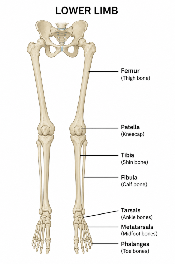
Femur(1) or thighbone, is the longest, heaviest, and strongest bone in the body. The femur and pelvis form the hip joint at the proximal end.
Patella(1) or kneecap, is a triangular bone that lies anterior to the knee joint. The patella is embedded in the tendon of the femoral extensors (quadriceps). It improves knee extension by reducing friction.
Tibia(1) or shinbone, is a large bone of the leg that is located directly below the knee. The tibia articulates with the femur at its proximal end, with the fibula and the tarsal bones at its distal end. It is the second largest bone in the human body and is responsible for transmitting the weight of the body from the femur to the foot.
Fibula(1) or calf bone, parallels and articulates with the tibia. It does not articulate with the femur and does not bear weight. The fibula acts as a site for muscle attachment and forms the lateral part of the ankle joint.
Tarsals(7) are the seven bones of the ankle. The ankle transmits the weight of the body from the tibia and the fibula to the foot.
Metatarsals(5) are the five bones of the foot.
Phalange(14) are the 14 bones of the leg finger. Each finger consists of three phalanges, except for the big toe that has only two.
Function of appendicular skeleton:-
The appendicular skeleton has two main functions.
1. voluntary movement:- The bones,muscles and joints of the limbs are involved in movement of the skeleton.
2.Protection of blood vessels and nerves:- These delicate structure along the length of bones of the limbs and are protected from injury by the associated muscles and skin .
3.Support and stability:- The girdles support the limbs and connect them to axial skeleton.
4.Weight bearing:- The lower limbs carry the body’s weight and help maintain balance and posture.
5.Attacjment for muscles:- Many musscles responsible for movement are attached to appendicular bones