


Introduction:-
It is a process by which the cells or tissues are fixed in chemical and partly physical state so that they can withstand subsequent treatment with various reagents, with minimal distortion of morphology and no decomposition
OBJECTIVES:-
After reading this lesson, you will be able to:
*state the aims of fixation *explain the principle of fixation *describe the properties and factors affecting fixation *explain types of fixation.
AIMS OF FIXATION:-
(c) To prevent tissues from changing their shape and size during processing (d) To harden the tissues (e) To allow clear staining of sections subsequently (f) To improve the optical differentiation of cells & tissue.
PRINCIPLE OF FIXATION:-
fixatives have a property of forming cross links between proteins, thereby forming a gel, keeping everything in their in vivo relation to each other.
AFFECTING FIXATION:-
weight of the fixative 3. pH of fixatives – Satisfactory fixation occurs between pH 6 and 8. Outside
this range, alteration in structure of cell may take place. 4. Temperature – Room temperature is alright for fixation. At high temperature
there may be distortion of tissues. 5. Volume changes – Cell volume changes because of the membrane
permeability and inhibition of respiration. 6. An ideal fixative should be cheap, nontoxic and non-inflammable. The
tissues may be kept in the fixative for a long time.
*Perfusion fixation
*Vapour fixation
*Coating/Spray fixation
*Freeze drying
*Microwave fixation/Stabilization
an excess of fixative. For all practical purposes immersion fixatives are most
useful. These may be divided into routine and special.
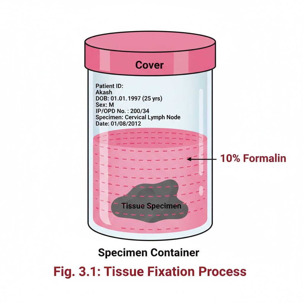
SIMPLE FIXATIVES:-
weight, called as formalin. Formaldehyde is commonly used as 4% solution,
giving 10% formalin for tissue fixation. Formalin is most commonly used
fixative. It is cheap, penetrates rapidly and does not over- harden the tissues.
The primary action of formalin is to form additive compounds with proteins
without precipitation. Formalin brings about fixation by converting the free
amine groups to methylene derivatives
If formalin is kept standing for a long time, a large amount of formic acid
is formed due to oxidation of formaldehyde and this tends to form artefact which is seen as brown pigment in the tissues. To avoid this buffered
formalin is used.
2. Absolute alcohol – it may be used as a fixative as it coagulates protein.
Due to its dehydrating property it removes water too fast from the tissues
and produces shrinkage of cells and distortion of morphology. It penetrates
slowly and over-hardens the tissues.
4. Mercuric chloride:-
It is a protein precipitant. However it causes great shrinkage of tissues hence seldom used alone. It gives brown colour to the tissues which needs to be removed by treatment with Iodine during
dehydration.
5. Potassium dichromate:-
6. Osmic acid :-
It is used for fixation of fatty tissues and nerves.
8. Osmium tetraoxide:-
9. Picric acid –
It precipitates proteins and combines with them to form picrates. Owing to its explosive nature when dry; it must be kept under a layer of water. Tissue fixed in picric acid also require thorough washing with water to remove colour. Tissue can not be kept in picric acid more than 24 hrs.
COMPOUND FIXATIVES:-
be removed in sections before staining by treatment with picric alcohol or
10% alcoholic solution of sodium hydroxide. The formation of this pigment
can be prevented by neutralizing or buffering the formal saline.
instead of acetic acid.
marrow, spleen & kidney.
of testicular biopsies.
specimen. The specimen should be completely submerged. 4. Special fixatives are used for preserving particular tissues. 5. Formalin vapours cause throat/ eye irritation hence mask/ eye glasses and
gloves should be used. 6. Tissues should be well fixed before dehydration. 7. Penetration of fixatives takes some time. It is necessary that the bigger
specimen should be given cuts so that the central part does not remain
unfixed.
8. Mercury pigment must be removed with Lugol’s iodine. 9. Biopsies cannot be kept for more than 24 hours in bouin’s fluid without
changing the alcohol. 10. Glutaraldehyde and osmion tetraoxide are used as fixatives for electron microscopy.