


INTRODUCTION:-
A microtome (from the Greek mikros, meaning “small”, and temnein, meaning “to cut”) is a tool used to cut extremely thin slices of material, known as sections.
MICROTOME:-
Various types of microtomes are available. Most commonly used microtome for routine histopathology is rotary microtome.
The most common applications of microtomes are:-
Traditional Histology Technique:-
Tissues are hardened by replacing water with paraffin. The tissue is then cut in the microtome at thicknesses varying from 2 to 50 µm. From there the tissue can be mounted on a microscope slide, stained with appropriate aqueous dye(s) after prior removal of the paraffin, and examined using a light microscope.
Cryosectioning Technique:-
Water-rich tissues are hardened by freezing and cut in the frozen state with a freezing microtome or microtome-cryostat; sections are stained and examined with a light microscope. This technique is much faster than traditional histology (15 minutes vs 16 hours) and is used in conjunction with medical procedures to achieve a quick diagnosis. Cryosections can also be used in immuno histochemistry as freezing tissue stops degradation of tissue faster than using a fixative and does not alter or mask its chemical composition as much.
Electron Microscopy Technique:-
After embedding tissues in epoxy resin, a microtome equipped with a glass or gem grade diamond knife is used to cut very thin sections (typically 60 to 100 nanometer). Sections are stained with an aqueous solution of an appropriate heavy metal salt and examined with a transmission electron microscope (TEM). This instrument is often called an ultramicrotome. The ultramicrotome is also used with its glass knife or an industrial grade diamond knife to cut survey sections prior to thin sectioning. These sections are of 0.5 to 1 µm thickness and are mounted on a glass slide and stained to locate areas of interest under a light microscope prior to thin sectioning for the TEM. Thin sectioning for the TEM is often done with a gem quality diamond knife.
Botanical Microtomy Technique:-
Hard materials like wood, bone and leather require a sledge microtome. These microtomes have heavier blades and cannot cut as thin as a regular microtome.
Rotary Mictrotome:-
It is most commonly used microtome. This device operates with a staged rotary action such that the actual cutting is part of the rotary motion. In a rotary microtome, the knife is typically fixed in a horizontal position A rotary action of the hand wheel actuate the cutting movement. Here the advantage over the rocking type is that it is heavier and there by more stable. Hard tissues can be cut without vibration. Serial sections or ribbons of sections can easily be obtained. The block holder or block (depends upon the type of cassette) is mounted on the steel carriage that moves up and down and is advanced by a micrometer screw.
Auto-cut microtome has built in motor drive with foot and hand control. With suitable accessories the machine can cut thin sections of paraffin wax blocks and 0.5 to 2.0 micrometer thin resin sections.
Advantages:-
1. The machine is heavy, so it is stable and does not vibrate during cutting.
2. Serial sections can be obtained.
3. Cutting angle and knife angle can be adjusted.
4. It may also be used for cutting celloidin embedded sections with the help
of special holder to set the knife.
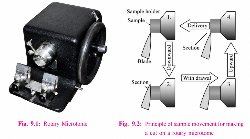
In the figure to the left, the principle of the cut is explained. Through the motion of the sample holder, the sample is cut by the knife position 1 to position 2), at which point the fresh section remains on the knife. At the highest point of the rotary motion, the sample holder is advanced by the same thickness as the section that is to be made, allowing for the next section to be made.
The flywheel in microtomes can be operated by hand. This has the advantage that a clean cut can be made, as the relatively large mass of the flywheel prevents the sample from being stopped during the sample cut. The flywheel in newer models is often integrated inside the microtome casing. The typical cut thickness for a rotary microtome is between 1 and 60 µm. For hard materials, such as a sample embedded in a synthetic resin, this design of microtome can allow for good “Semi-thin” sections with a thickness of as low as 0.5 µm.
Sledge Microtome:-
it is a device where the sample is placed into a fixed holder (shuttle), the sledge placed upon a linear bearing, a design that allows for the microtome to readily cut many coarse sections. Applications for this design of microtome are of the preparation of large samples, such as those embedded in paraffin for biological preparations. Typical cut thickness achievable on a sledge microtome is between is 10 and 60 micron.
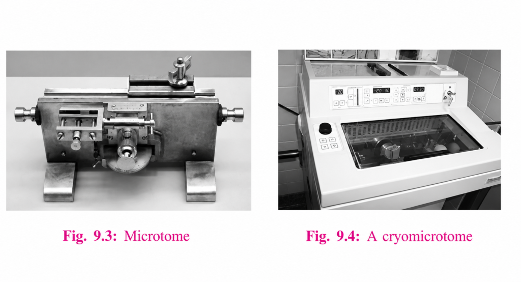
Cryomicrotome:-
For the cutting of frozen samples, many rotary microtomes can be adapted to cut in a liquid nitrogen chamber, in a so-called cryomicrotome setup. The
reduced temperature allows for the hardness of the sample to be increased, such as by undergoing a glass transition, which allows for the preparation of semi
thin samples. However the sample temperature and the knife temperature must be controlled in order to optimise the resultant sample thickness.
Ultramicrotome:-
A ribbon of ultrathin sections prepared by room temperature ultramicrotomy, floating on water in the boat of a diamond knife used to cut the sections. The
knife blade is the edge at the upper end of the trough of water.
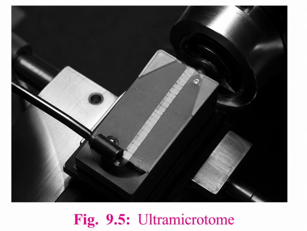
An ultramicrotome is a main tool of ultramicrotomy. It can allow for the preparation of extremely thin sections, with the device functioning in the same manner as a rotational microtome, but with very tight tolerances on the mechanical construction. As a result of the careful mechanical construction, the linear thermal expansion of the mounting is used to provide very fine control of the thickness.
These extremely thin cuts are important for use with transmission electron microscope (TEM) and Serial Block-Face Scanning Electron Microscopy
(SBFSEM), and are sometimes also important for light-optical microscopy. The typical thickness of these cuts is between 40 and 100 nm for transmission
electron microscopy and often between 30 and 50 nm for SBFSEM. Thicker sections up to 500 nm thick are also taken for specialized TEM applications or
for light microscopy survey sections to select an area for the final thin sections. Diamond knives (preferably) and glass knives are used with ultramicrotomes.
To collect the sections they are floated on top of a liquid as they are cut and are carefully picked up onto grids suitable for TEM specimen viewing. The
thickness of the section can be estimated by the thin-film interference colors of reflected light that are seen as a result of the extremely low sample thickness.
Vibrating microtome:-
The vibrating microtome operates by cutting using a vibrating blade, allowing the resultant cut to be made with less pressure than would be required for a stationary blade. The vibrating microtome is usually used for difficult biological samples. The cut thickness is usually around 30-500 µm for live tissue and 10-500 µm for fixed tissue.
Saw microtome:-
The saw microtome is especially for hard materials such as teeth or bones. The microtome of this type has a recessed rotating saw, which slices through the
sample. The minimal cut thickness is approximately 30 µm, and can be made for comparatively large samples.
Laser microtome:- A conceptual diagram of laser microtome operation
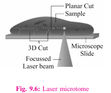
The laser microtome is an instrument for contact free slicing. Prior preparation of the sample through embedding, freezing or chemical fixation is not required, thereby minimizing the artifacts from preparation methods. Alternately this design of microtome can also be used for very hard materials, such as bones or teeth as well as some ceramics. Dependent upon the properties of the sample material, the thickness achievable is between 10 and 100 µm.
The device operates using a cutting action of an infra-red laser. As the laser emits a radiation in the near infra-red, in this wavelength regime the laser can interact with biological materials. Through sharp focusing of the probe within the sample, a focal point of very high intensity, up to TW/cm2, can be achieved.
Through the non-linear interaction of the optical penetration in the focal region a material separation in a process known as photo-disruption is introduced. By
limiting the laser pulse durations to the femtoseconds range, the energy.
expended at the target region is precisely controlled, thereby limiting the interaction zone of the cut to under a micrometre. External to this zone the ultra
short beam application time introduces minimal to no thermal damage to the remainder of the sample.
The laser radiation is directed onto a fast scanning mirror based optical system which allows for three dimensional positioning of the beam crossover, whilst
allowing for beam traversal to the desired region of interest. The combination of high power with a high raster rate allows the scanner to cut large areas of
sample in a short time. In the laser microtome the laser-microdissection of internal areas in tissues, cellular structures, and other types of small features is
also possible.
MICROTOME KNIFE:-
manually or by the use of automatic machine.
Honing
Stropping –
expensive and used for ultramicrotomy.
1. Low profile blade – Usually used for cutting small and soft biopsies like
kidney and liver biopsies.
2. High profile blade-Used for any tissue like myometrium, breast tumor or
skin.
2. Resistant to both corrosion and heat.
3. Hardness of blade can be compared with the steel knife.
2. Disposable blades are not as rigid as steel knife:
Care of the Microtome Knife:-
*The knife should be cleaned with xylene before and after use.
*When knife is being stored for a long time, it should be smeared with grease
or good grade of light oil.
*Knife edge should not be touched.
*Knife edge should never be come badly nicked. It is advisable to use
separate knife for cutting hard issue like bone.
*The above points are important if re usable knife is being used.
1 For routine histopathology rotary microtome is used.
2 Ultramicrotome is used to cut semi-thin sections or ultrathin sections.
3 Traditional type of knife requires honing and stropping to smoothen the
cutting edge.
4 Disposable knives are expensive but do not need honing or stropping.
5 Knife edge is spoiled if properly decalcified tissue is not used.