


Prussian Blue Staining:-
Aim: To demonstrate the presence of iron in tissues.
Principle: The ferric iron in tissue combines with potassium ferro cyanide to form ferric-ferro cyanide. This compound has bright blue color (prussian blue). Prussian blue precipitate is insoluble, hence it can be combined with other staining methods.
Control: Hemosiderin positive tissue
Reagents:-
1. 2%Hydrochloric acid
concentrated hydrochloric acid 2ml
Distilled water 98ml
2. 2% potassium ferrocyanide
Potassium ferrocyanide 2mg
Distilled water 100ml
3. 0.15% Basic fuchsin
Basic fuchsin 0.15g
50% ethyl alcohol 100ml
Procedure:-
1. Bring section to water.
2. Mix equal volume of 2% potassium ferrocyanide and 2% hydrochloric acid.
Pour the solution on the slide and keep it for 20 minutes.
3. Wash thoroughly with water.
4. Counter-stain with basic fuchsin or eosin for 30 seconds.
5. Wash with water, dehydrate, clear in xylene and mount in DPX.
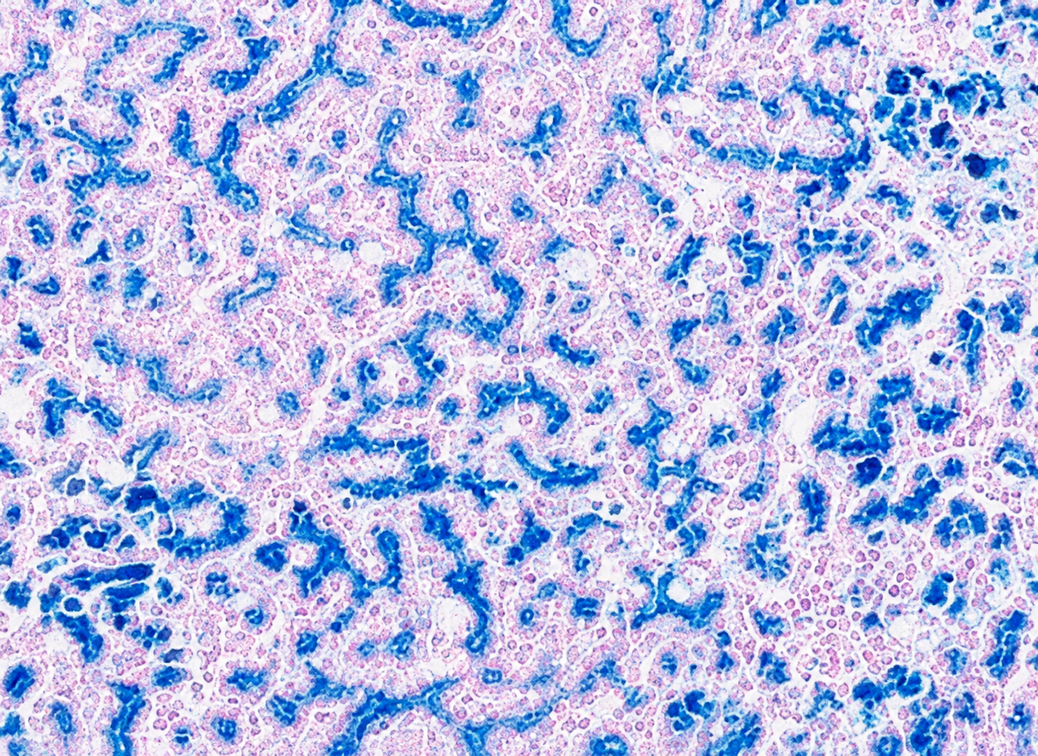
Result:-
Ferric iron blue
Nuclei red
Other tissues shades of pink
Note: All traces of ferrocyanide should be removed before it is counter-stained,
otherwise a dark red fine precipitate will form.