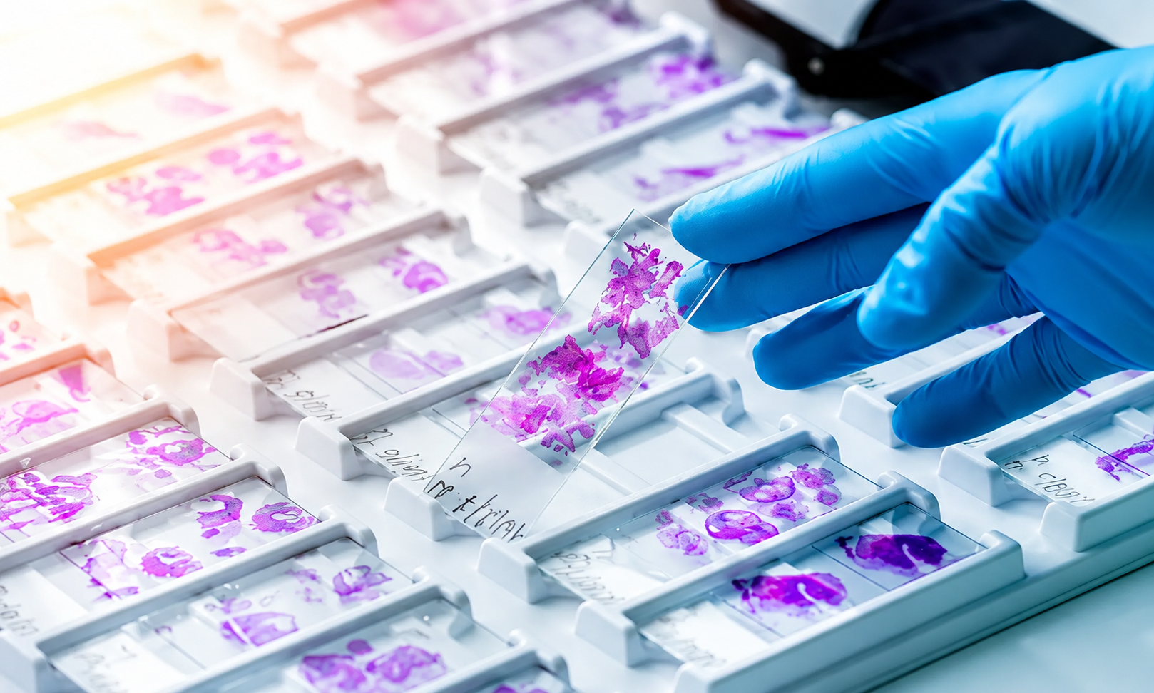


Introduction:-
Some pathological specimens require special handling and need to be processed in a different way to reach the final diagnosis. Examples include eyeball, bones and bone marrow biopsy. The technical person needs to be aware of these special specimens so that appropriate measures can be taken before the grossing procedures are undertaken.
BONE:-
Normal human skeleton has two main types of bones: cortical or compact bone which is hard, solid and very strong and forms shafts of long bones i.e. the femur and tibia etc; and spongy or trabecular/ cancellous bone is found in the marrow cavities and is a mesh of bone strands which is almost ideal weight bearing structure particularly in the femoral head and vertebrae. The three major components of bone are mineral, cells and an organic extra-cellular matrix i.e. collagen fibers. The main bulk of bone is approximately 70% mineral and 30% organic components by weight. Bone cells are relatively few as opposed to marrow cells. The mineral of bone is mainly calcium and phosphate.
Techniques for the demonstration of bone and its components include:-
* For decalcified bone: frozen, paraffin, or celloidin sections, transmission electron microscopy.
* For mineralized bone: frozen, plastic, scanning and transmission electron microscopy.
* For mineralized bone: frozen, plastic, scanning and transmission electron microscopy.
In order to obtain satisfactory paraffin sections of bone, inorganic calcium must be removed from the organic collagen matrix, calcified cartilage and surrounding tissues. This is called decalcification and is carried out by chemical agents, either with acids to form soluble calcium salts or with chelating agents that bind to calcium ions. Any acid, however well buffered, can have damaging effects on tissue staining. The problem increases with acidity of solution and duration of decalcification period. It mostly affects the nuclei which fail to take up hematoxylin and other basic dyes. These effects can be reduced by doing the
decalcification end point test, post-decalcification acid removal and adjustment of the stain procedure.
decalcification end point test, post-decalcification acid removal and adjustment of the stain procedure.
DECALCIFYING AGENTS:-
Acids:-
Acid decalcifiers can be divided into two groups: strong (inorganic) and weak.
Strong Inorganic acids:-
Acids:-
Acid decalcifiers can be divided into two groups: strong (inorganic) and weak.
Strong Inorganic acids:-
e.g. nitric and hydrochloric acids may be used as simple aqueous solutions (5-10%). They decalcify rapidly but cause tissue swelling and can seriously damage tissue stainability if used longer than 24-48 hours. Old nitric acid is particularly damaging and should be replaced with fresh stock They also damage tissue antigens for immunohistochemistry and enzymes may be totally lost. They can be used for small needle biopsies to permit rapid diagnosis within 24hrs. They can also be used for large or heavily mineralized bones with decalcification progress being carefully monitored.
Aqueous Nitric acid, 5-10%
Nitric acid 5-10 ml
Distilled water To make 100ml
Formalin-nitric acid:-
Formaldehyde (37-40%) 10 ml
Distilled water 80 ml
Distilled water To make 100ml
Formalin-nitric acid:-
Formaldehyde (37-40%) 10 ml
Distilled water 80 ml
Nitric acid 10 ml
Weak organic acids:-
e.g. fromic, acetic and picric acid. Of these three formic acid is the only weak acid which is used in decalcification. Other two are used as components of other fixatives. Formic acid solutions can be aqueous (5-10%), buffered or combined with formalin. The formalin-10% formic acid mixture fixes and decalcifies simultaneously and can be used for small biopsies. Formic acid is suitable for most routine surgical specimens, particularly when immunohistochemistry is needed. Decalcification is usually complete in 1-10 days and decalcification progress should be monitered by a decalcification end point test.
Aqueous formic acid:-
90% stock formic acid 5-10 ml
Distilled water To make 100 ml
Distilled water To make 100 ml
Formic acid-formalin:-
90% stock formic acid 5-10 ml
Formaldehyde (37-40%) 5 ml
Distilled water To make 100 ml
Formaldehyde (37-40%) 5 ml
Distilled water To make 100 ml
Buffered formic acid:-
20% aqueous sodium citrate 65 ml
90% stock formic acid 35 ml
90% stock formic acid 35 ml
CHELATING AGENTS:-
The chelating agent used for decalcification is ethylene-diaminetetracetic acid (EDTA). Although called an acid, it does not act like acids. EDTA will not bind to calcium below pH 3 and is faster at pH 7-7.4. This is a very slow process that does not damage tissues or their stainability, it also retains good antigen preservation for immunohistochemistry or enzyme staining and electron microscopy. The time required to totally decalcify dense cortical bone may be 6-8 weeks or longer although small bone spicules may be decalcified in less than a week.
Formalin-EDTA:-
EDTA, disodium salt 5.5 g
Distilled water 90 ml
Formaldehyde (37-40% stock) 10 ml
Distilled water 90 ml
Formaldehyde (37-40% stock) 10 ml
Aqueous EDTA, pH 7.0-7.4:-
EDTA, disodium salt 250 g
Distilled water 1750 ml
Distilled water 1750 ml
If solution is cloudy, adjust to pH 7 with sodium hydroxide.
Factors influencing the rate of decalcification:-
There are several factors influencing the rate of decalcification. The concentration and volume of the active reagent, including the temperature at which the reaction takes place, are important at all times. The more concentrated acids solutions decalcify bone more rapidly but are more harmful to the tissue. The usuall recommended ratio of volume of decalifying fluid to volume of tissue is 20:1 and the fluid should be changed several times during the decalcification.
Process:-
Increased temperature accelerates decalcification, but it also increases the tissue damage and loss of heat sensitive antigen and enzymes. Other factors that contribute include the age of patient, type of bone, size of specimen and solution agitation. Mature cortical bone decalcifies more slowly than immature
bone.
Treatment following decalcification:-
Acids can be removed from tissues or neutralized chemically after decalcification is complete. It can be done by immersing the bone into either saturated lithium carbonate solution or 5-10% aqueous sodium bicarbonate solution for several hours. Many laboratories rinse the specimens with running tap water for a period of time.
Eyeball:-
The eye should be placed in fixative as soon as practical after removal. Many of the tissues, the retina in particular, are very sensitive to anoxia and the longer you wait to fix the eye, the greater will be the artifacts, making interpretation difficult.
If eyes arrive in formalin and have been fixed for 2 days, wash them in water to remove the formalin (2 changes about 5 minutes each) and place them in enough 50% ethanol to cover the eye. Let the eye equilibrate overnight. Change the alcohol the next day and equilibrate for a second day. The eye should return to a normal volume and should not be indented or shrunken. For sectioning the eye it is best to wait 2 days with the eye in 50% ethanol.
If eyes arrive in formalin and have been fixed for 2 days, wash them in water to remove the formalin (2 changes about 5 minutes each) and place them in enough 50% ethanol to cover the eye. Let the eye equilibrate overnight. Change the alcohol the next day and equilibrate for a second day. The eye should return to a normal volume and should not be indented or shrunken. For sectioning the eye it is best to wait 2 days with the eye in 50% ethanol.