


INTRODUCTION:-
Nucleoproteins are combinations of basic proteins and nucleic acids. The two nucleic acids are deoxyribonucleic acid (DNA), which is mainly found in nucleus and ribonucleic acid (RNA) which is located in the cytoplasm of cells, mainly in the ribosomes. Both DNA and RNA molecules consist of alternate sugar and phosphate groups with a nitrogenous base being attached to each sugar group. The sugar in DNA is deoxyribose and in RNA it is ribose. The demonstration of nucleic acid depends upon either the reaction of dyes with the phosphate groups or the production of aldehydes from the sugars.
DNA:-The demonstration of DNA is either by Feulgen technique (which demonstrates the sugar deoxyribose) or the methyl green-pyronin technique (where the phosphates combine with basic dye methyl green at acidic pH). It can also be
demonstrated by fluorescent methods using acridine orange, but is considered less reliable than the above mentioned methods. The definitive and most
sensitive technique is in situ hybridization.
FEULGEN TECHNIQUE:-
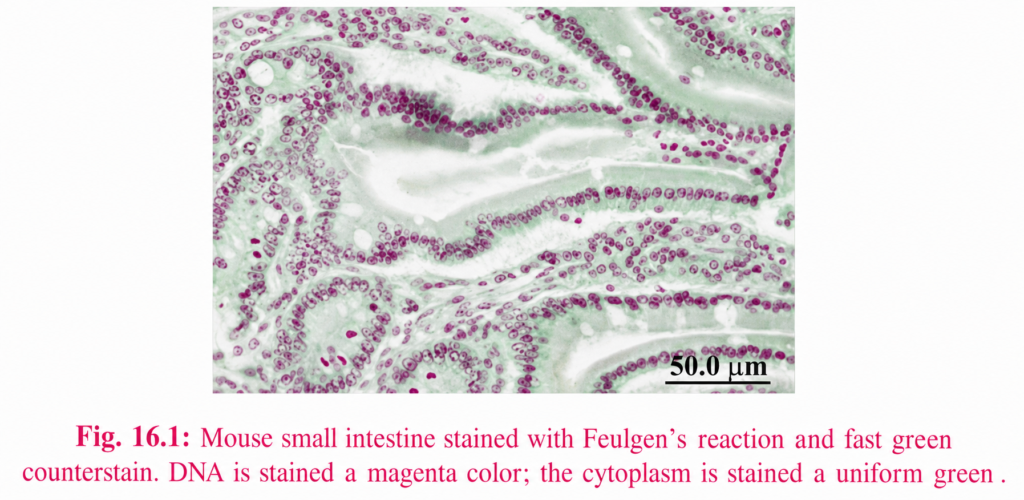
This technique involves mild acid hydrolysis with 1M hydrochloric acid at 60°C to break the purine-deoxyribose bond, the resulting exposed aldehydes are then reacted with Schiff’s reagent to stain the DNA red-purple in color.
Feulgen nuclear reaction for DNA:- Fixation: Not critical but do not use Bouin’s fixative.
Solutions:-
(a) 1 M hydrochloric acid
Hydrochloric acid (conc.) 8.5 ml
Distilled water 91.5 ml
(b) Schiff reagent
(c) Bisulfite solution
10% potassium metabisulfite 5 ml
1M hydrochloric acid 5 ml
Distilled water 100 ml
1. Bring all sections to water.Rinse 2.sections in 1M HCl at room temperature.
3. Place sections in 1M HCl at 60°C
4.Rinse in 1M HCl at room temperature, 1 minute.
5. Transfer sections to Schiff’s reagent, 45 minutes.
6. Rinse sections in bisulfate solution, 2 minutes, repeating twice again.
7. Rinse well in distilled water.
8. Counterstain if required in 1% light green, 2 minutes.
9. Wash in water.
10. Dehydrate through alcohols to xylene and mount.
Results:-
DNA red-purple
Cytoplasm green
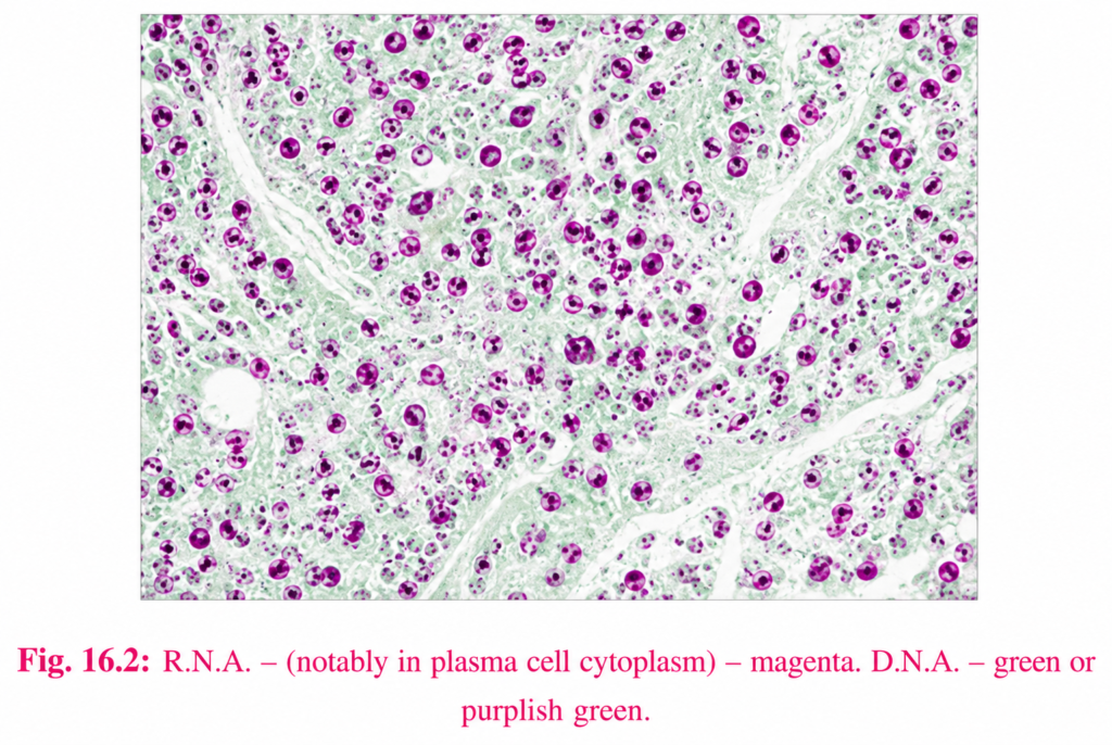
RNA:-The method of choice for demonstrating RNA is the methyl green-pyronin
technique.
Methyl green-pyronin:-Methyl green is an impure dye containing methyl violet. When methyl violet has
been removed by washing with chloroform, the pure methyl green appears and
is specific for DNA. Both dyes are cationinc, when used in combination methyl
green binds preferentially and specifically to DNA, and pyronin binds RNA.
Methyl green-pyroninmethod for RNA
Fixation: Carnoy preferred, but formalin acceptable.
Staining Solution: Methyl green pyronin Y
2% methyl green (chloroform washed) 9ml
2% pyronin Y 4 ml
Acetate buffer pH 4.8 23 ml
Glycerol 14 m
Mix well before use.
Method:-
1. Take sections down to water.
2. Rinse in acetate buffer pH 4.8.
3. Place in methyl green-pyronin Y solution for 25 min.
4. Rinse in buffer.5. Blot dry.
6. Rinse in 93% ethanol, then in absolute ethanol.
7. Rinse in xylene and mount.
Results:-
DNA green-blue
RNA red
MITOCHONDRIA:-Mitochondria are the cytoplasmic organelle found in variable numbers in all
animal cells. Large number of mitochondria in the cells can change the
appearance of cells. Mitochondria are considered the ‘power houses’ of the cell
as many of the energy producing biochemical reactions like oxidative
phosphorylation and Krebs cycle activity takes place in mitochondria.
Mitochondria can be demonstrated by electron microscopy, enzyme histochemistry
and histological methods however electron microscopy is the most satisfactory
method. Histopathological methods such as Altman’s technique for mitochondria
is simple and useful for demonstration of mitochondria.
Altman’s technique for mitochondria:-
Fixation:-Champy’s fluid is usually recommended, Helly’s fluid works equally as well.
Aniline-acid fuchsin – saturated solution of acid fuchsin in 5% aniline in
distilled water.
Differentiator 1
Saturated alcohalic picric acid 10 ml
30% alcohol 40 ml
Differentiator 2
Saturated alcohalic picric acid 5 ml
30% alcohol 40 ml
Method:-
1. Take sections down to water.
2. Flood sections with aniline-acid fuchsin solution.
3. Gently heat the slide until steam rises and leave for 5 min.
4. Rinse in tap water.
5. Differentiate in solution 1 until the excess red stain is removed.
6. Completely differentiate in solution 2, controlling microscopically.
7. Dehydrate rapidly in two changes of absolute alcohal.
8. Clear in xylene and mount in DPX.
Results:-
Mitochondria red
RBC and nuclei red
Background tissue yellow