


Verheoff stain for collagen:-
Aim:– To identify collagen and elastic tissue in the same section.
Principle: In the presence of ferric salts (oxidizers) elastic fibers stain with
hematoxylin, along with the nuclei.
Control: skin
Reagents
1. Verhoeff’s solution: Freshly prepared solution gives best result.
Solution A
Hematoxylin 5 gm
Absolute alcohol 100ml
Dissolve hematoxylin with the aid of heat, cool and filter
Solution BFerric chloride 10gm
Distilled water 100ml
Solution C
Iodine 2gm
Potassium iodide 4gm
Distilled water 100ml
Add 8ml of solution B into 20ml of solution A and then add 8ml of
solution C
2. 2% Ferric chloride solution
3. 1% aqueous solution of acid fuchsin
4. Saturated aqueous solution of picric acid
5. Van Gieson’s stain
Acid Fuchsin 1% (aqueous) 5ml
Saturated aqueous solution of picric acid 100ml
6. Sodium thiosulphate, 5% (aqueous solution)
Procedure
1. Deparaffinize and take the section to water.
2. Stain in Verhoeff solution until the section is black.
3. Wash in distilled water.
4. Differentiate in 2% Ferric chloride with agitation for few minutes. Check differentiation by rinsing in distilled water. Under the microscope the elastic fibers and nuclei should stain black and rest of the tissue should be light grey.
5. Put in 5% sodium thiosulphate for 1 minute.
6. Wash in tap water for 5minutes.
7. Counter-stain with Van Gieson’s stain for 1-2 minutes.
8. Differentiate in 95% alcohol.
9. Dehydrate in absolute alcohol two times.
10. Clear in xylene and mount in DPX.
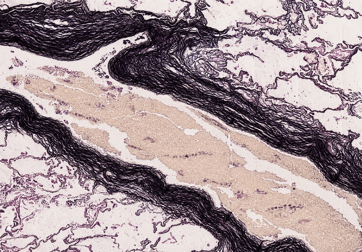
Result:-
Elastic fibres black
Nuclei black
Collagen red
Other tissues yellow
Note: It is a rapid method but fails to demonstrate fine fibers