


STAINING METHODS TO DEMONSTRATE SPECIAL/ SPECIFIC TISSUES:-
Introduction:-
Biological tissue has little inherent contrast in either the light or electron microscope. Staining is employed to give both contrast to the tissue as well as highlighting particular features of interest. Where the underlying mechanistic chemistry of staining is understood, the term histochemistry is used.
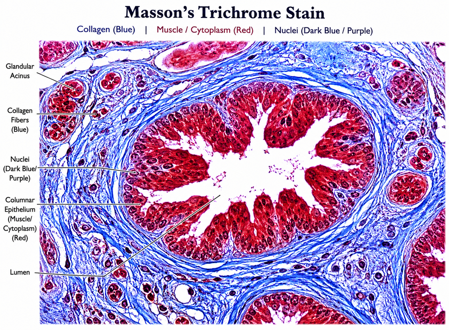
TRICHROME STAIN:-
A combination of three different dyes is used to identify different cells and tissue elements.
Aim:-To identify the collagen and muscle fibers in a histological section.
Reagents:-
1. Bouin’s solution
Saturated picric acid 75 ml
Formaldehyde (37-40%) 25 ml
Glacial acetic acid 5 ml
*Mix all the reagents well. This solution improves the trichrome stain
quality.
2. Weigert’s iron hematoxylin stock solution
Stock solution A
Hematoxylin 1gm
95% alcohol 100 ml
Stock solution B
29% Ferric chloride in water 4ml
Distilled water 100 ml Hydrochloric acid, concentrated 1.0 ml
3. Weigert’s iron hematoxylin working solution – Mix equal parts of solution A and B (This solution works for three months.)
4. Biebrich scarlet acid fuchsin solution.
1% Biebric Scarlet-Acid Fuchsin solution (aqueous solution) 90 ml
1% Acid Fuchsin (Aqueous) 10 ml
1% Glacial acitic acid 1 ml
5. Phosphomolybdic acid-Phosphotungstic Acid Solution.
5% Phosphomolybdic Acid 25ml
5%phosphotungstic Acid 25ml
6. Aniline blue solution
Aniline blue solution 2.5gm
Glacial acitic acid 2ml
Distilled water 100 ml
Control: skin.
Procedure:-
1. De-paraffinize and rehydrate through graded alcohol.
2. Wash in distilled water.
3. Fix the slides in Bouin’s solution for one hour at 560C.
4. Rinse in running tap water for 5 to 10 minutes to remove yellow color.
5. Stain in Weigert’s Iron Hematoxylin solution for 10 minutes.
6. Rinse in warm tap water for 10 minutes.
7. Wash in distilled water.
8. Put Biebric Scarlet Acid Fuchsin solution for 10 to 15 minutes.
9. Wash in distilled water.
10. Differentiate in Phosphomolybdic-Phosphotungstic Acid solution for 10 to
15 minutes.
11. Put the sections in Aniline blue solution for 5-10 minutes.
12. Rinse in distilled water briefly.
13. Differentiate in acetic acid solution for 2-5 minutes.
14. Wash in distilled water.
15. Dehydrate quickly through 95% alcohol and absolute alcohol. (These steps will wipe off Biebric Scarlet acid Fuchsin staining)
16. Clear in xyline and mount in DPX.
Result:-
Glycogen, muscle fibre and keratin red
Collagen and bone blue/green
Nuclei brown/black
Note: This stain can be used on frozen sections also.